The carbohydrate recognition domain of Langerin reveals high structural similarity with the one of DC-SIGN but an additional, calcium-independent sugar-binding site.
Chatwell, L., Holla, A., Kaufer, B.B., Skerra, A.(2008) Mol Immunol 45: 1981-1994
- PubMed: 18061677
- DOI: https://doi.org/10.1016/j.molimm.2007.10.030
- Primary Citation of Related Structures:
3P7F, 3P7G, 3P7H - PubMed Abstract:
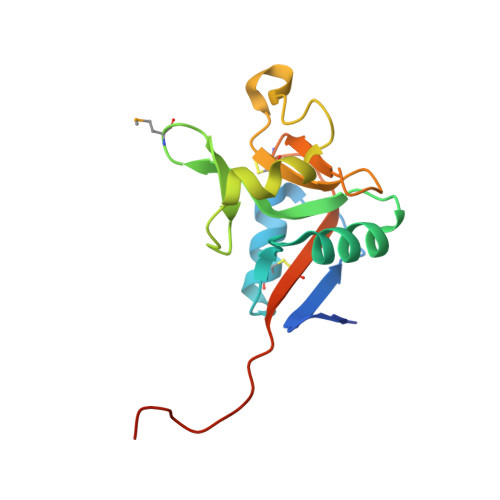
Langerin is a type II transmembrane oligosaccharide receptor on Langerhans cells (LCs), a prominent subclass of dendritic cells (DCs) that mediate immune responses in epithelia and play a role in HIV degradation. Its extracellular moiety comprises a neck region with several heptad repeats and an exposed carboxy-terminal calcium-type carbohydrate-recognition domain (CRD). The CRD of human Langerin, which was expressed as a soluble protein in the periplasm of E. coli, was crystallized both alone and in the presence of two sugars, followed by X-ray analyses to resolutions of 2.5A for apo-Langerin and to 1.6A and 2.1A for the complexes with mannose and maltose, respectively. The fold of the Langerin CRD (dubbed LangA) resembles that of other typical C-type lectins such as DC-SIGN. However, especially in the long loop region (LLR), which is responsible for carbohydrate-binding, two additional secondary structure elements are present: a 3(10) helix and a small beta-sheet arising from the extended beta-strand 2, which enters into a hairpin and a new strand beta2'. Unexpectedly, the crystal structures in the presence of maltose and mannose reveal two sugar-binding sites. One is calcium-dependent and structurally conserved in the C-type lectin family whereas the second one represents a novel, calcium-independent type. Based on these data, a model for the binding of mannan, a component of many endogenous as well as viral glycoproteins, is proposed and the differences in binding behavior between Langerin and DC-SIGN with respect to the Lewis X carbohydrate antigen and its derivatives can be explained. Therefore, the crystal structure of LangA should be helpful for the development of new marker reagents selective for LCs and also of therapeutic compounds that may enhance the inhibitory role of Langerin towards HIV infection.
Organizational Affiliation:
Lehrstuhl für Biologische Chemie, Technische Universität München, 85350 Freising-Weihenstephan, Germany.