


Crystal structure of H.pylori phosphopantetheine adenylyltransferase mutant I4V/N76Y
Chen, C.H., Cheng, C.S., Yin, H.S.To be published.
Experimental Data Snapshot
wwPDB Validation 3D Report Full Report

Entity ID: 1 | |||||
|---|---|---|---|---|---|
| Molecule | Chains | Sequence Length | Organism | Details | Image |
| Phosphopantetheine adenylyltransferase | 157 | Helicobacter pylori 26695 | Mutation(s): 2 Gene Names: coaD, kdtB, HP_1475 EC: 2.7.7.3 | 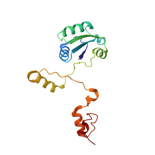 | |
UniProt | |||||
Find proteins for O26010 (Helicobacter pylori (strain ATCC 700392 / 26695)) Explore O26010 Go to UniProtKB: O26010 | |||||
Entity Groups | |||||
| Sequence Clusters | 30% Identity50% Identity70% Identity90% Identity95% Identity100% Identity | ||||
| UniProt Group | O26010 | ||||
Sequence AnnotationsExpand | |||||
| |||||
| Ligands 2 Unique | |||||
|---|---|---|---|---|---|
| ID | Chains | Name / Formula / InChI Key | 2D Diagram | 3D Interactions | |
| SO4 Query on SO4 | B [auth A], C [auth A], D [auth A], E [auth A] | SULFATE ION O4 S QAOWNCQODCNURD-UHFFFAOYSA-L |  | ||
| ACY Query on ACY | F [auth A] | ACETIC ACID C2 H4 O2 QTBSBXVTEAMEQO-UHFFFAOYSA-N |  | ||
| Length ( Å ) | Angle ( ˚ ) |
|---|---|
| a = 54.022 | α = 90 |
| b = 69.459 | β = 90 |
| c = 95.92 | γ = 90 |
| Software Name | Purpose |
|---|---|
| REFMAC | refinement |
| PDB_EXTRACT | data extraction |
| HKL-2000 | data collection |
| HKL-2000 | data reduction |
| HKL-2000 | data scaling |
| MOLREP | phasing |
RCSB PDB (citation) is hosted by
RCSB PDB is a member of the

