3NBN

Crystal structure of a dimer of Notch Transcription Complex trimers on HES1 DNA


Experimental Data Snapshot

  • Method: X-RAY DIFFRACTION
  • Resolution: 3.45 Å
  • R-Value Free: 0.298 
  • R-Value Work: 0.254 
  • R-Value Observed: 0.256 

wwPDB Validation   3D Report Full Report


This is version 1.3 of the entry. See complete history


Literature

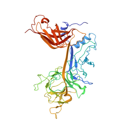
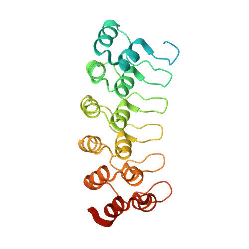
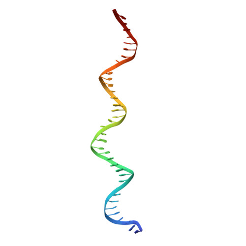
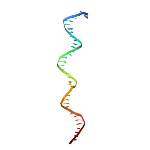
Structural and mechanistic insights into cooperative assembly of dimeric Notch transcription complexes.

Arnett, K.L.Hass, M.McArthur, D.G.Ilagan, M.X.Aster, J.C.Kopan, R.Blacklow, S.C.

(2010) Nat Struct Mol Biol 17: 1312-1317

  • DOI: https://doi.org/10.1038/nsmb.1938
  • Primary Citation of Related Structures:  
    3NBN

  • PubMed Abstract: 

    Ligand-induced proteolysis of Notch produces an intracellular effector domain that transduces essential signals by regulating the transcription of target genes. This function relies on the formation of transcriptional activation complexes that include intracellular Notch, a Mastermind co-activator and the transcription factor CSL bound to cognate DNA. These complexes form higher-order assemblies on paired, head-to-head CSL recognition sites. Here we report the X-ray structure of a dimeric human Notch1 transcription complex loaded on the paired site from the human HES1 promoter. The small interface between the Notch ankyrin domains could accommodate DNA bending and untwisting to allow a range of spacer lengths between the two sites. Cooperative dimerization occurred on the human and mouse Hes5 promoters at a sequence that diverged from the CSL-binding consensus at one of the sites. These studies reveal how promoter organizational features control cooperativity and, thus, the responsiveness of different promoters to Notch signaling.


  • Organizational Affiliation

    Department of Cancer Biology, Dana-Farber Cancer Institute, Boston, Massachusetts, USA.


Macromolecules

Find similar proteins by:  (by identity cutoff)  |  3D Structure
Entity ID: 1
MoleculeChains Sequence LengthOrganismDetailsImage
Recombining binding protein suppressor of hairless
A, D
433Homo sapiensMutation(s): 0 
Gene Names: IGKJRBIGKJRB1RBPJRBPJKRBPSUH
UniProt & NIH Common Fund Data Resources
Find proteins for Q06330 (Homo sapiens)
Explore Q06330 
Go to UniProtKB:  Q06330
PHAROS:  Q06330
GTEx:  ENSG00000168214 
Entity Groups  
Sequence Clusters30% Identity50% Identity70% Identity90% Identity95% Identity100% Identity
UniProt GroupQ06330
Sequence Annotations
Expand
  • Reference Sequence
Find similar proteins by:  (by identity cutoff)  |  3D Structure
Entity ID: 2
MoleculeChains Sequence LengthOrganismDetailsImage
Neurogenic locus notch homolog protein 1
B, E
256Homo sapiensMutation(s): 0 
Gene Names: NOTCH1TAN1
UniProt & NIH Common Fund Data Resources
Find proteins for P46531 (Homo sapiens)
Explore P46531 
Go to UniProtKB:  P46531
PHAROS:  P46531
GTEx:  ENSG00000148400 
Entity Groups  
Sequence Clusters30% Identity50% Identity70% Identity90% Identity95% Identity100% Identity
UniProt GroupP46531
Sequence Annotations
Expand
  • Reference Sequence
Find similar proteins by:  (by identity cutoff)  |  3D Structure
Entity ID: 3
MoleculeChains Sequence LengthOrganismDetailsImage
Mastermind-like protein 1
C, F
63Homo sapiensMutation(s): 0 
Gene Names: KIAA0200MAML1
UniProt & NIH Common Fund Data Resources
Find proteins for Q92585 (Homo sapiens)
Explore Q92585 
Go to UniProtKB:  Q92585
PHAROS:  Q92585
GTEx:  ENSG00000161021 
Entity Groups  
Sequence Clusters30% Identity50% Identity70% Identity90% Identity95% Identity100% Identity
UniProt GroupQ92585
Sequence Annotations
Expand
  • Reference Sequence
Find similar nucleic acids by:  (by identity cutoff)  |  3D Structure
Entity ID: 4
MoleculeChains LengthOrganismImage
DNA, HES1 promoterG [auth X]37N/A
Sequence Annotations
Expand
  • Reference Sequence
Find similar nucleic acids by:  (by identity cutoff)  |  3D Structure
Entity ID: 5
MoleculeChains LengthOrganismImage
DNA, HES1 promoterH [auth Y]37N/A
Sequence Annotations
Expand
  • Reference Sequence
Experimental Data & Validation

Experimental Data

  • Method: X-RAY DIFFRACTION
  • Resolution: 3.45 Å
  • R-Value Free: 0.298 
  • R-Value Work: 0.254 
  • R-Value Observed: 0.256 
  • Space Group: C 1 2 1
Unit Cell:
Length ( Å )Angle ( ˚ )
a = 295.112α = 90
b = 108.059β = 102.52
c = 87.239γ = 90
Software Package:
Software NamePurpose
DENZOdata reduction
SCALEPACKdata scaling
MOLREPphasing
DMphasing
REFMACrefinement
PDB_EXTRACTdata extraction
ADSCdata collection
HKL-2000data reduction
HKL-2000data scaling

Structure Validation

View Full Validation Report


Entry History 

Deposition Data

Revision History  (Full details and data files)

  • Version 1.0: 2010-11-03
    Type: Initial release
  • Version 1.1: 2011-07-13
    Changes: Version format compliance
  • Version 1.2: 2017-11-08
    Changes: Refinement description
  • Version 1.3: 2023-09-06
    Changes: Data collection, Database references, Refinement description