3N9D

Monoclinic Structure of P. aeruginosa LigD phosphoesterase domain


Experimental Data Snapshot

  • Method: X-RAY DIFFRACTION
  • Resolution: 2.30 Å
  • R-Value Free: 0.263 
  • R-Value Work: 0.212 
  • R-Value Observed: 0.217 

wwPDB Validation   3D Report Full Report


This is version 1.2 of the entry. See complete history


Literature

Structure of bacterial LigD 3'-phosphoesterase unveils a DNA repair superfamily

Nair, P.A.Smith, P.Shuman, S.

(2010) Proc Natl Acad Sci U S A 107: 12822-12827

  • DOI: https://doi.org/10.1073/pnas.1005830107
  • Primary Citation of Related Structures:  
    3N9B, 3N9D

  • PubMed Abstract: 

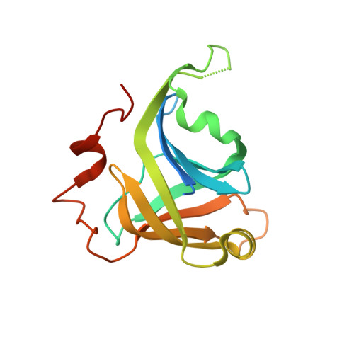
    The DNA ligase D (LigD) 3'-phosphoesterase (PE) module is a conserved component of the bacterial nonhomologous end-joining (NHEJ) apparatus that performs 3' end-healing reactions at DNA double-strand breaks. Here we report the 1.9 A crystal structure of Pseudomonas aeruginosa PE, which reveals that PE exemplifies a unique class of DNA repair enzyme. PE has a distinctive fold in which an eight stranded beta barrel with a hydrophobic interior supports a crescent-shaped hydrophilic active site on its outer surface. Six essential side chains coordinate manganese and a sulfate mimetic of the scissile phosphate. The PE active site and mechanism are unique vis à vis other end-healing enzymes. We find PE homologs in archaeal and eukaryal proteomes, signifying that PEs comprise a DNA repair superfamily.


  • Organizational Affiliation

    Molecular Biology Program, Sloan-Kettering Institute, New York, NY 10065, USA.


Macromolecules
Find similar proteins by:  (by identity cutoff)  |  3D Structure
Entity ID: 1
MoleculeChains Sequence LengthOrganismDetailsImage
Probable ATP-dependent DNA ligase171Pseudomonas aeruginosaMutation(s): 0 
Gene Names: NP_250828.1PA2138
UniProt
Find proteins for Q9I1X7 (Pseudomonas aeruginosa (strain ATCC 15692 / DSM 22644 / CIP 104116 / JCM 14847 / LMG 12228 / 1C / PRS 101 / PAO1))
Explore Q9I1X7 
Go to UniProtKB:  Q9I1X7
Entity Groups  
Sequence Clusters30% Identity50% Identity70% Identity90% Identity95% Identity100% Identity
UniProt GroupQ9I1X7
Sequence Annotations
Expand
  • Reference Sequence
Experimental Data & Validation

Experimental Data

  • Method: X-RAY DIFFRACTION
  • Resolution: 2.30 Å
  • R-Value Free: 0.263 
  • R-Value Work: 0.212 
  • R-Value Observed: 0.217 
  • Space Group: C 1 2 1
Unit Cell:
Length ( Å )Angle ( ˚ )
a = 90.997α = 90
b = 56.585β = 118.33
c = 43.503γ = 90
Software Package:
Software NamePurpose
HKL-2000data collection
PHASERphasing
PHENIXrefinement
HKL-2000data reduction
SCALEPACKdata scaling

Structure Validation

View Full Validation Report


Entry History 

Deposition Data

Revision History  (Full details and data files)

  • Version 1.0: 2010-08-11
    Type: Initial release
  • Version 1.1: 2011-07-13
    Changes: Version format compliance
  • Version 1.2: 2023-09-06
    Changes: Data collection, Database references, Derived calculations, Refinement description