3LSM

Pyranose 2-oxidase H167A mutant with flavin N(5) sulfite adduct


Experimental Data Snapshot

  • Method: X-RAY DIFFRACTION
  • Resolution: 1.70 Å
  • R-Value Free: 0.195 
  • R-Value Work: 0.160 
  • R-Value Observed: 0.160 

wwPDB Validation   3D Report Full Report


Ligand Structure Quality Assessment 


This is version 1.5 of the entry. See complete history


Literature

H-bonding and positive charge at the N5/O4 locus are critical for covalent flavin attachment in trametes pyranose 2-oxidase.

Tan, T.C.Pitsawong, W.Wongnate, T.Spadiut, O.Haltrich, D.Chaiyen, P.Divne, C.

(2010) J Mol Biol 402: 578-594

  • DOI: https://doi.org/10.1016/j.jmb.2010.08.011
  • Primary Citation of Related Structures:  
    3LSH, 3LSI, 3LSK, 3LSM

  • PubMed Abstract: 

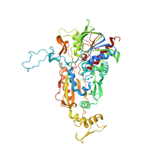
    Flavoenzymes perform a wide range of redox reactions in nature, and a subclass of flavoenzymes carry covalently bound cofactor. The enzyme-flavin bond helps to increase the flavin's redox potential to facilitate substrate oxidation in several oxidases. The formation of the enzyme-flavin covalent bond--the flavinylation reaction--has been studied for the past 40 years. For the most advocated mechanism of autocatalytic flavinylation, the quinone methide mechanism, appropriate stabilization of developing negative charges at the flavin N(1) and N(5) loci is crucial. Whereas the structural basis for stabilization at N(1) is relatively well studied, the structural requisites for charge stabilization at N(5) remain less clear. Here, we show that flavinylation of histidine 167 of pyranose 2-oxidase from Trametes multicolor requires hydrogen bonding at the flavin N(5)/O(4) locus, which is offered by the side chain of Thr169 when the enzyme is in its closed, but not open, state. Moreover, our data show that additional stabilization at N(5) by histidine 548 is required to ensure high occupancy of the histidyl-flavin bond. The combination of structural and spectral data on pyranose 2-oxidase mutants supports the quinone methide mechanism. Our results demonstrate an elaborate structural fine-tuning of the active site to complete its own formation that couples efficient holoenzyme synthesis to conformational substates of the substrate-recognition loop and concerted movements of side chains near the flavinylation ligand.


  • Organizational Affiliation

    Division of Biochemistry, School of Biotechnology, Royal Institute of Technology, Albanova University Center, Roslagstullsbacken 21, Stockholm, Sweden.


Macromolecules
Find similar proteins by:  (by identity cutoff)  |  3D Structure
Entity ID: 1
MoleculeChains Sequence LengthOrganismDetailsImage
Pyranose 2-oxidase
A, B
623Trametes ochraceaMutation(s): 1 
Gene Names: p2o
EC: 1.1.3.10
UniProt
Find proteins for Q7ZA32 (Trametes ochracea)
Explore Q7ZA32 
Go to UniProtKB:  Q7ZA32
Entity Groups  
Sequence Clusters30% Identity50% Identity70% Identity90% Identity95% Identity100% Identity
UniProt GroupQ7ZA32
Sequence Annotations
Expand
  • Reference Sequence
Small Molecules
Ligands 4 Unique
IDChains Name / Formula / InChI Key2D Diagram3D Interactions
SFD
Query on SFD

Download Ideal Coordinates CCD File 
C [auth A],
H [auth B]
(S)-10-((2S,3S,4R)-5-((S)-((S)-(((2R,3S,4R,5R)-5-(6-AMINO-9H-PURIN-9-YL)-3,4-DIHYDROXY-TETRAHYDROFURAN-2-YL)METHOXY)(HYDROXY)PHOSPHORYLOXY)(HYDROXY)PHOSPHORYLOXY)-2,3,4-TRIHYDROXYPENTYL)-7,8-DIMETHYL-2,4-DIOXO-2,3,4,4A-TETRAHYDROBENZO[G]PTERIDINE-5(10H)-SULFONIC ACID
C27 H35 N9 O18 P2 S
WCBOBURWKHIISA-LDVRFEIFSA-N
12P
Query on 12P

Download Ideal Coordinates CCD File 
G [auth A],
K [auth B]
DODECAETHYLENE GLYCOL
C24 H50 O13
WRZXKWFJEFFURH-UHFFFAOYSA-N
GLC
Query on GLC

Download Ideal Coordinates CCD File 
F [auth A]alpha-D-glucopyranose
C6 H12 O6
WQZGKKKJIJFFOK-DVKNGEFBSA-N
SO3
Query on SO3

Download Ideal Coordinates CCD File 
D [auth A],
E [auth A],
I [auth B],
J [auth B]
SULFITE ION
O3 S
LSNNMFCWUKXFEE-UHFFFAOYSA-L
Experimental Data & Validation

Experimental Data

  • Method: X-RAY DIFFRACTION
  • Resolution: 1.70 Å
  • R-Value Free: 0.195 
  • R-Value Work: 0.160 
  • R-Value Observed: 0.160 
  • Space Group: P 43 21 2
Unit Cell:
Length ( Å )Angle ( ˚ )
a = 101.568α = 90
b = 101.568β = 90
c = 249.577γ = 90
Software Package:
Software NamePurpose
MAR345data collection
FFTmodel building
CCP4model building
REFMACrefinement
XDSdata reduction
XSCALEdata scaling
FFTphasing
CCP4phasing

Structure Validation

View Full Validation Report


Ligand Structure Quality Assessment 


Entry History 

Deposition Data

Revision History  (Full details and data files)

  • Version 1.0: 2010-08-25
    Type: Initial release
  • Version 1.1: 2011-07-13
    Changes: Version format compliance
  • Version 1.2: 2017-11-01
    Changes: Data collection, Refinement description
  • Version 1.3: 2020-07-29
    Type: Remediation
    Reason: Carbohydrate remediation
    Changes: Advisory, Data collection, Derived calculations, Structure summary
  • Version 1.4: 2021-10-13
    Changes: Database references, Structure summary
  • Version 1.5: 2024-02-21
    Changes: Data collection