3LCD

Inhibitor Bound to A DFG-In structure of the Kinase Domain of CSF-1R


Experimental Data Snapshot

  • Method: X-RAY DIFFRACTION
  • Resolution: 2.50 Å
  • R-Value Free: 0.265 
  • R-Value Work: 0.216 
  • R-Value Observed: 0.218 

wwPDB Validation   3D Report Full Report


Ligand Structure Quality Assessment 


This is version 1.2 of the entry. See complete history


Literature

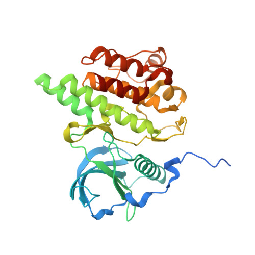
Structure-based drug design enables conversion of a DFG-in binding CSF-1R kinase inhibitor to a DFG-out binding mode

Meyers, M.J.Pelc, M.Kamtekar, S.Day, J.Poda, G.I.Hall, M.K.Michener, M.L.Reitz, B.A.Mathis, K.J.Pierce, B.S.Parikh, M.D.Mischke, D.A.Long, S.A.Parlow, J.J.Anderson, D.R.Thorarensen, A.

(2010) Bioorg Med Chem Lett 20: 1543-1547

  • DOI: https://doi.org/10.1016/j.bmcl.2010.01.078
  • Primary Citation of Related Structures:  
    3LCD, 3LCO

  • PubMed Abstract: 

    The work described herein demonstrates the utility of structure-based drug design (SBDD) in shifting the binding mode of an HTS hit from a DFG-in to a DFG-out binding mode resulting in a class of novel potent CSF-1R kinase inhibitors suitable for lead development.


  • Organizational Affiliation

    Pfizer Global Research & Development, St. Louis Laboratories, 700 Chesterfield Parkway West, Chesterfield, MO 63017, United States. marvin.j.meyers@pfizer.com


Macromolecules
Find similar proteins by:  (by identity cutoff)  |  3D Structure
Entity ID: 1
MoleculeChains Sequence LengthOrganismDetailsImage
Macrophage colony-stimulating factor 1 receptor329Homo sapiensMutation(s): 0 
Gene Names: CSF-1RCSF1RFMS
EC: 2.7.10.1
UniProt & NIH Common Fund Data Resources
Find proteins for P07333 (Homo sapiens)
Explore P07333 
Go to UniProtKB:  P07333
PHAROS:  P07333
GTEx:  ENSG00000182578 
Entity Groups  
Sequence Clusters30% Identity50% Identity70% Identity90% Identity95% Identity100% Identity
UniProt GroupP07333
Sequence Annotations
Expand
  • Reference Sequence
Small Molecules
Ligands 2 Unique
IDChains Name / Formula / InChI Key2D Diagram3D Interactions
BDY
Query on BDY

Download Ideal Coordinates CCD File 
D [auth A]N~3~-(2,6-dichlorobenzyl)-5-(4-{[(2R)-2-(pyrrolidin-1-ylmethyl)pyrrolidin-1-yl]carbonyl}phenyl)pyrazine-2,3-diamine
C27 H30 Cl2 N6 O
PVBCEWZVPUKFNT-HXUWFJFHSA-N
SO4
Query on SO4

Download Ideal Coordinates CCD File 
B [auth A],
C [auth A]
SULFATE ION
O4 S
QAOWNCQODCNURD-UHFFFAOYSA-L
Binding Affinity Annotations 
IDSourceBinding Affinity
BDY Binding MOAD:  3LCD IC50: 40 (nM) from 1 assay(s)
PDBBind:  3LCD IC50: 40 (nM) from 1 assay(s)
Experimental Data & Validation

Experimental Data

  • Method: X-RAY DIFFRACTION
  • Resolution: 2.50 Å
  • R-Value Free: 0.265 
  • R-Value Work: 0.216 
  • R-Value Observed: 0.218 
  • Space Group: P 21 21 21
Unit Cell:
Length ( Å )Angle ( ˚ )
a = 57.649α = 90
b = 74.125β = 90
c = 91.024γ = 90
Software Package:
Software NamePurpose
HKL-2000data collection
PHASERphasing
REFMACrefinement
HKL-2000data reduction
HKL-2000data scaling

Structure Validation

View Full Validation Report


Ligand Structure Quality Assessment 


Entry History 

Deposition Data

Revision History  (Full details and data files)

  • Version 1.0: 2010-03-02
    Type: Initial release
  • Version 1.1: 2011-07-13
    Changes: Advisory, Version format compliance
  • Version 1.2: 2017-07-26
    Changes: Data collection, Refinement description, Source and taxonomy