


Structural determination and tryptophan fluorescence of heterokaryon incompatibility C2 protein (HET-C2), a fungal glycolipid transfer protein (GLTP), provide novel insights into glycolipid specificity and membrane interaction by the GLTP fold.
Kenoth, R., Simanshu, D.K., Kamlekar, R.K., Pike, H.M., Molotkovsky, J.G., Benson, L.M., Bergen, H.R., Prendergast, F.G., Malinina, L., Venyaminov, S.Y., Patel, D.J., Brown, R.E.(2010) J Biol Chem 285: 13066-13078
- PubMed: 20164530
- DOI: https://doi.org/10.1074/jbc.M109.093203
- Primary Citation of Related Structures:
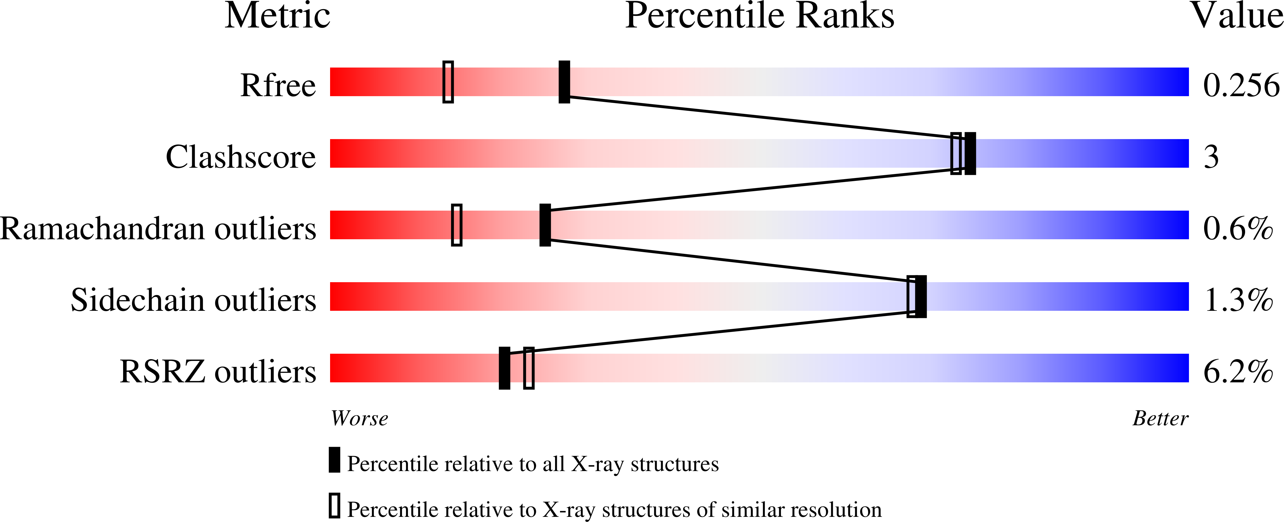
3KV0 - PubMed Abstract:
HET-C2 is a fungal protein that transfers glycosphingolipids between membranes and has limited sequence homology with human glycolipid transfer protein (GLTP). The human GLTP fold is unique among lipid binding/transfer proteins, defining the GLTP superfamily. Herein, GLTP fold formation by HET-C2, its glycolipid transfer specificity, and the functional role(s) of its two Trp residues have been investigated. X-ray diffraction (1.9 A) revealed a GLTP fold with all key sugar headgroup recognition residues (Asp(66), Asn(70), Lys(73), Trp(109), and His(147)) conserved and properly oriented for glycolipid binding. Far-UV CD showed secondary structure dominated by alpha-helices and a cooperative thermal unfolding transition of 49 degrees C, features consistent with a GLTP fold. Environmentally induced optical activity of Trp/Tyr/Phe (2:4:12) detected by near-UV CD was unaffected by membranes containing glycolipid but was slightly altered by membranes lacking glycolipid. Trp fluorescence was maximal at approximately 355 nm and accessible to aqueous quenchers, indicating free exposure to the aqueous milieu and consistent with surface localization of the two Trps. Interaction with membranes lacking glycolipid triggered significant decreases in Trp emission intensity but lesser than decreases induced by membranes containing glycolipid. Binding of glycolipid (confirmed by electrospray injection mass spectrometry) resulted in a blue-shifted emission wavelength maximum (approximately 6 nm) permitting determination of binding affinities. The unique positioning of Trp(208) at the HET-C2 C terminus revealed membrane-induced conformational changes that precede glycolipid uptake, whereas key differences in residues of the sugar headgroup recognition center accounted for altered glycolipid specificity and suggested evolutionary adaptation for the simpler glycosphingolipid compositions of filamentous fungi.
Organizational Affiliation:
Hormel Institute, University of Minnesota, Austin, Minnesota 55912, USA.


