High-resolution X-ray crystal structure of bovine H-protein at 0.88 A resolution
Higashiura, A., Kurakane, T., Matsuda, M., Suzuki, M., Inaka, K., Sato, M., Kobayashi, T., Tanaka, T., Tanaka, H., Fujiwara, K., Nakagawa, A.(2010) Acta Crystallogr D Biol Crystallogr 66: 698-708
- PubMed: 20516622
- DOI: https://doi.org/10.1107/S0907444910010668
- Primary Citation of Related Structures:
3KLR - PubMed Abstract:
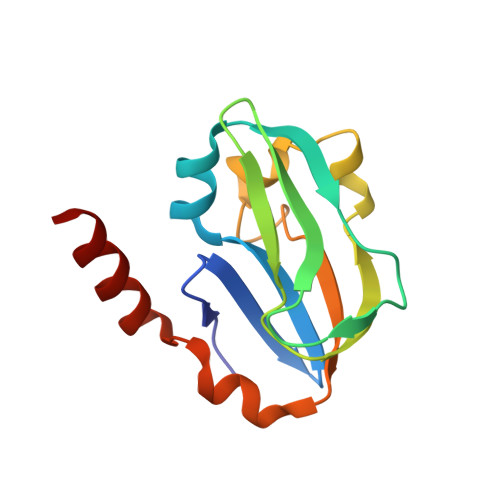
Recent technical improvements in macromolecular X-ray crystallography have significantly improved the resolution limit of protein structures. However, examples of high-resolution structure determination are still limited. In this study, the X-ray crystal structure of bovine H-protein, a component of the glycine cleavage system, was determined at 0.88 A resolution. This is the first ultrahigh-resolution structure of an H-protein. The data were collected using synchrotron radiation. Because of limitations of the hardware, especially the dynamic range of the CCD detector, three data sets (high-, medium- and low-resolution data sets) were measured in order to obtain a complete set of data. To improve the quality of the merged data, the reference data set was optimized for merging and the merged data were assessed by comparing merging statistics and R factors against the final model and the number of visualized H atoms. In addition, the advantages of merging three data sets were evaluated. The omission of low-resolution reflections had an adverse effect on visualization of H atoms in hydrogen-omit maps. Visualization of hydrogen electron density is a good indicator for assessing the quality of high-resolution X-ray diffraction data.
Organizational Affiliation:
Institute for Protein Research, Osaka University, Japan.