3JU0

Structure of the arm-type binding domain of HAI7 integrase


Experimental Data Snapshot

  • Method: X-RAY DIFFRACTION
  • Resolution: 1.60 Å
  • R-Value Free: 0.212 
  • R-Value Work: 0.148 
  • R-Value Observed: 0.151 

wwPDB Validation   3D Report Full Report


This is version 1.4 of the entry. See complete history


Literature

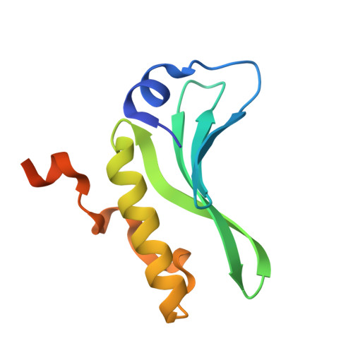
Structures of the arm-type binding domains of HPI and HAI7 integrases

Szwagierczak, A.Antonenka, U.Popowicz, G.M.Sitar, T.Holak, T.A.Rakin, A.

(2009) J Biol Chem 284: 31664-31671

  • DOI: https://doi.org/10.1074/jbc.M109.059261
  • Primary Citation of Related Structures:  
    3JTZ, 3JU0

  • PubMed Abstract: 

    The structures of the N-terminal domains of two integrases of closely related but not identical asn tDNA-associated genomic islands, Yersinia HPI (high pathogenicity island; encoding siderophore yersiniabactin biosynthesis and transport) and an Erwinia carotovora genomic island with yet unknown function, HAI7, have been resolved. Both integrases utilize a novel four-stranded beta-sheet DNA-binding motif, in contrast to the known proteins that bind their DNA targets by means of three-stranded beta-sheets. Moreover, the beta-sheets in Int(HPI) and Int(HAI7) are longer than those in other integrases, and the structured helical N terminus is positioned perpendicularly to the large C-terminal helix. These differences strongly support the proposal that the integrases of the genomic islands make up a distinct evolutionary branch of the site-specific recombinases that utilize a unique DNA-binding mechanism.


  • Organizational Affiliation

    Max Planck Institute for Biochemistry, D-82152 Martinsried, Germany.


Macromolecules
Find similar proteins by:  (by identity cutoff)  |  3D Structure
Entity ID: 1
MoleculeChains Sequence LengthOrganismDetailsImage
Phage integrase108Pectobacterium atrosepticumMutation(s): 0 
Gene Names: intBECA1679
UniProt
Find proteins for Q6D6K1 (Pectobacterium atrosepticum (strain SCRI 1043 / ATCC BAA-672))
Explore Q6D6K1 
Go to UniProtKB:  Q6D6K1
Entity Groups  
Sequence Clusters30% Identity50% Identity70% Identity90% Identity95% Identity100% Identity
UniProt GroupQ6D6K1
Sequence Annotations
Expand
  • Reference Sequence
Experimental Data & Validation

Experimental Data

  • Method: X-RAY DIFFRACTION
  • Resolution: 1.60 Å
  • R-Value Free: 0.212 
  • R-Value Work: 0.148 
  • R-Value Observed: 0.151 
  • Space Group: P 65
Unit Cell:
Length ( Å )Angle ( ˚ )
a = 52.53α = 90
b = 52.53β = 90
c = 79.47γ = 120
Software Package:
Software NamePurpose
MAR345data collection
MOLREPphasing
REFMACrefinement
XDSdata reduction
XSCALEdata scaling

Structure Validation

View Full Validation Report


Entry History 

Deposition Data

Revision History  (Full details and data files)

  • Version 1.0: 2009-10-06
    Type: Initial release
  • Version 1.1: 2011-07-13
    Changes: Version format compliance
  • Version 1.2: 2013-01-16
    Changes: Database references
  • Version 1.3: 2017-11-01
    Changes: Refinement description
  • Version 1.4: 2023-11-01
    Changes: Data collection, Database references, Refinement description