3INZ

H57T Hfq from Pseudomonas aeruginosa


Experimental Data Snapshot

  • Method: X-RAY DIFFRACTION
  • Resolution: 1.70 Å
  • R-Value Free: 0.218 
  • R-Value Work: 0.149 
  • R-Value Observed: 0.153 

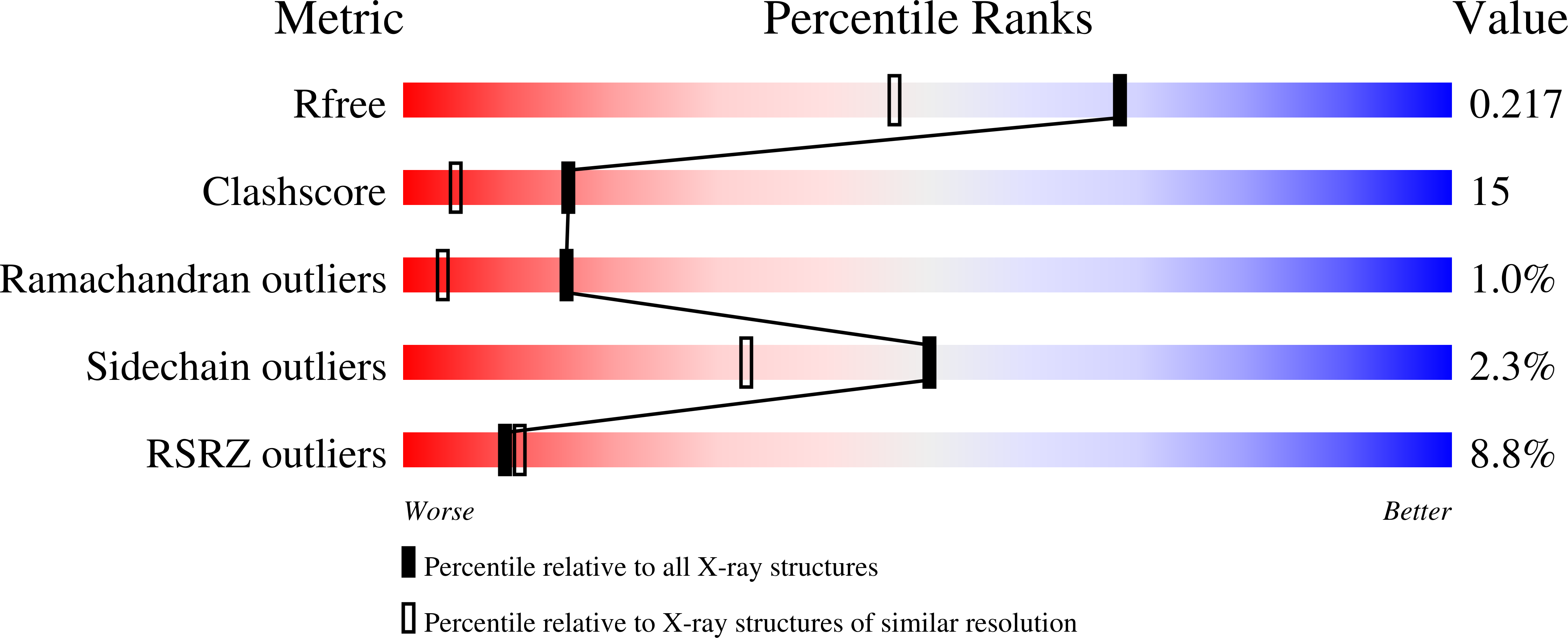
wwPDB Validation   3D Report Full Report


This is version 1.3 of the entry. See complete history


Literature

The structures of mutant forms of Hfq from Pseudomonas aeruginosa reveal the importance of the conserved His57 for the protein hexamer organization.

Moskaleva, O.Melnik, B.Gabdulkhakov, A.Garber, M.Nikonov, S.Stolboushkina, E.Nikulin, A.

(2010) Acta Crystallogr Sect F Struct Biol Cryst Commun 66: 760-764

  • DOI: https://doi.org/10.1107/S1744309110017331
  • Primary Citation of Related Structures:  
    3INZ, 3M4G

  • PubMed Abstract: 

    The bacterial Sm-like protein Hfq forms homohexamers both in solution and in crystals. The monomers are organized as a continuous beta-sheet passing through the whole hexamer ring with a common hydrophobic core. Analysis of the Pseudomonas aeruginosa Hfq (PaeHfq) hexamer structure suggested that solvent-inaccessible intermonomer hydrogen bonds created by conserved amino-acid residues should also stabilize the quaternary structure of the protein. In this work, one such conserved residue, His57, in PaeHfq was replaced by alanine, threonine or asparagine. The crystal structures of His57Thr and His57Ala Hfq were determined and the stabilities of all of the mutant forms and of the wild-type protein were measured. The results obtained demonstrate the great importance of solvent-inaccessible conserved hydrogen bonds between the Hfq monomers in stabilization of the hexamer structure.


  • Organizational Affiliation

    Institute of Protein Research, RAS, Russia.


Macromolecules
Find similar proteins by:  (by identity cutoff)  |  3D Structure
Entity ID: 1
MoleculeChains Sequence LengthOrganismDetailsImage
Protein hfq
A, B, C, D, E
A, B, C, D, E, F
82Pseudomonas aeruginosaMutation(s): 1 
Gene Names: hfqPA4944
UniProt
Find proteins for Q9HUM0 (Pseudomonas aeruginosa (strain ATCC 15692 / DSM 22644 / CIP 104116 / JCM 14847 / LMG 12228 / 1C / PRS 101 / PAO1))
Explore Q9HUM0 
Go to UniProtKB:  Q9HUM0
Entity Groups  
Sequence Clusters30% Identity50% Identity70% Identity90% Identity95% Identity100% Identity
UniProt GroupQ9HUM0
Sequence Annotations
Expand
  • Reference Sequence
Small Molecules
Ligands 3 Unique
IDChains Name / Formula / InChI Key2D Diagram3D Interactions
CD
Query on CD

Download Ideal Coordinates CCD File 
G [auth A]
H [auth B]
I [auth B]
K [auth C]
L [auth D]
G [auth A],
H [auth B],
I [auth B],
K [auth C],
L [auth D],
N [auth E],
O [auth F],
P [auth F],
Q [auth F]
CADMIUM ION
Cd
WLZRMCYVCSSEQC-UHFFFAOYSA-N
CL
Query on CL

Download Ideal Coordinates CCD File 
M [auth D]CHLORIDE ION
Cl
VEXZGXHMUGYJMC-UHFFFAOYSA-M
NA
Query on NA

Download Ideal Coordinates CCD File 
J [auth B]SODIUM ION
Na
FKNQFGJONOIPTF-UHFFFAOYSA-N
Experimental Data & Validation

Experimental Data

  • Method: X-RAY DIFFRACTION
  • Resolution: 1.70 Å
  • R-Value Free: 0.218 
  • R-Value Work: 0.149 
  • R-Value Observed: 0.153 
  • Space Group: P 21 21 21
Unit Cell:
Length ( Å )Angle ( ˚ )
a = 61.29α = 90
b = 71.16β = 90
c = 104.41γ = 90
Software Package:
Software NamePurpose
MAR345dtbdata collection
PHENIXmodel building
PHENIXrefinement
XDSdata reduction
XDSdata scaling
PHENIXphasing

Structure Validation

View Full Validation Report


Entry History 

Deposition Data

Revision History  (Full details and data files)

  • Version 1.0: 2010-08-04
    Type: Initial release
  • Version 1.1: 2011-07-13
    Changes: Version format compliance
  • Version 1.2: 2021-10-13
    Changes: Data collection, Database references, Derived calculations, Structure summary
  • Version 1.3: 2023-09-06
    Changes: Data collection, Refinement description