3INS

STRUCTURE OF INSULIN. RESULTS OF JOINT NEUTRON AND X-RAY REFINEMENT


Experimental Data Snapshot

  • Method: X-RAY DIFFRACTION
  • Resolution: 1.50 Å
  • R-Value Work: 0.182 

  • Method: NEUTRON DIFFRACTION
  • Resolution: 2.20 Å

wwPDB Validation   3D Report Full Report


This is version 1.3 of the entry. See complete history


Literature

Structure of insulin: results of joint neutron and X-ray refinement.

Wlodawer, A.Savage, H.Dodson, G.

(1989) Acta Crystallogr B 45: 99-107

  • DOI: https://doi.org/10.1107/s0108768188011012
  • Primary Citation of Related Structures:  
    3INS

  • PubMed Abstract: 

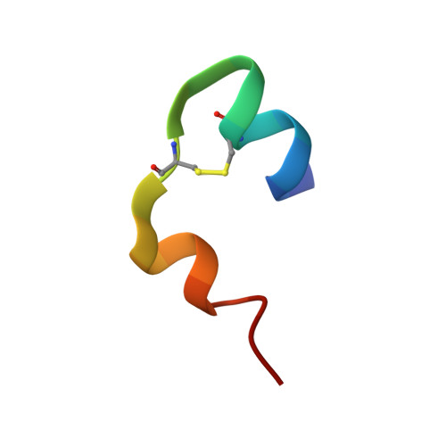
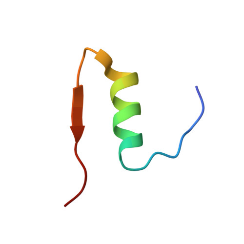
    Neutron diffraction data for porcine 2Zn insulin were collected to 2.2 A resolution from a single crystal deuterated by slow exchange of mother liquor. A joint neutron/X-ray restrained-least-squares refinement was undertaken using the neutron data, as well as the 1.5 A resolution X-ray data collected previously. The final R factors were 0.182 for the X-ray data and 0.191 for the neutron data. Resulting atomic coordinates were compared with the initial X-ray model, showing a total r.m.s. shift of 0.36 A for the protein and 0.6 A for the solvent. Protonation of a number of individual amino acids was investigated by analysis of the neutron maps. No D atoms were found between the carboxylates of Glu B13 which make an intermolecular contact, suggesting nonbonded interaction rather than the predicted hydrogen bond. Amide hydrogen exchange was investigated in a refinement of their atomic occupancies. Regions of unexchanged amide groups were found in the center of the B helices. The results of this study emphasize the limited amount of information available in neutron diffraction studies of proteins at resolution lower than 2 A.


  • Organizational Affiliation

    Center for Chemical Physics, National Bureau of Standards, Gaithersburg, MD 20899.


Macromolecules

Find similar proteins by:  Sequence   |   3D Structure  

Entity ID: 1
MoleculeChains Sequence LengthOrganismDetailsImage
INSULIN (CHAIN A)
A, C
21Sus scrofaMutation(s): 0 
UniProt
Find proteins for P01315 (Sus scrofa)
Explore P01315 
Go to UniProtKB:  P01315
Entity Groups  
Sequence Clusters30% Identity50% Identity70% Identity90% Identity95% Identity100% Identity
UniProt GroupP01315
Sequence Annotations
Expand
  • Reference Sequence
Find similar proteins by:  (by identity cutoff)  |  3D Structure
Entity ID: 2
MoleculeChains Sequence LengthOrganismDetailsImage
INSULIN (CHAIN B)
B, D
30Sus scrofaMutation(s): 0 
UniProt
Find proteins for P01315 (Sus scrofa)
Explore P01315 
Go to UniProtKB:  P01315
Entity Groups  
Sequence Clusters30% Identity50% Identity70% Identity90% Identity95% Identity100% Identity
UniProt GroupP01315
Sequence Annotations
Expand
  • Reference Sequence
Small Molecules
Ligands 1 Unique
IDChains Name / Formula / InChI Key2D Diagram3D Interactions
ZN
Query on ZN

Download Ideal Coordinates CCD File 
E [auth B],
F [auth D]
ZINC ION
Zn
PTFCDOFLOPIGGS-UHFFFAOYSA-N
Experimental Data & Validation

Experimental Data

  • Method: X-RAY DIFFRACTION
  • Resolution: 1.50 Å
  • R-Value Work: 0.182 
  • Space Group: H 3
  • Method: NEUTRON DIFFRACTION
  • Resolution: 2.20 Å
  • Space Group: H 3
Unit Cell:
Length ( Å )Angle ( ˚ )
a = 82.5α = 90
b = 82.5β = 90
c = 34γ = 120
Software Package:
Software NamePurpose
PROLSQrefinement

Structure Validation

View Full Validation Report


Entry History 

Deposition Data

Revision History  (Full details and data files)

  • Version 1.0: 1989-01-09
    Type: Initial release
  • Version 1.1: 2008-03-03
    Changes: Version format compliance
  • Version 1.2: 2011-07-13
    Changes: Data collection, Non-polymer description, Version format compliance
  • Version 1.3: 2017-11-29
    Changes: Derived calculations, Other