Crystallographic and Molecular Dynamics Analysis of Loop Motions Unmasking the Peptidoglycan-Binding Site in Stator Protein MotB of Flagellar Motor
Reboul, C.F., Andrews, D.A., Nahar, M.F., Buckle, A.M., Roujeinikova, A.(2011) PLoS One 6: e18981-e18981
- PubMed: 21533052
- DOI: https://doi.org/10.1371/journal.pone.0018981
- Primary Citation of Related Structures:
3IMP - PubMed Abstract:
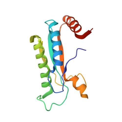
The C-terminal domain of MotB (MotB-C) shows high sequence similarity to outer membrane protein A and related peptidoglycan (PG)-binding proteins. It is believed to anchor the power-generating MotA/MotB stator unit of the bacterial flagellar motor to the peptidoglycan layer of the cell wall. We previously reported the first crystal structure of this domain and made a puzzling observation that all conserved residues that are thought to be essential for PG recognition are buried and inaccessible in the crystal structure. In this study, we tested a hypothesis that peptidoglycan binding is preceded by, or accompanied by, some structural reorganization that exposes the key conserved residues. We determined the structure of a new crystalline form (Form B) of Helicobacter pylori MotB-C. Comparisons with the existing Form A revealed conformational variations in the petal-like loops around the carbohydrate binding site near one end of the β-sheet. These variations are thought to reflect natural flexibility at this site required for insertion into the peptidoglycan mesh. In order to understand the nature of this flexibility we have performed molecular dynamics simulations of the MotB-C dimer. The results are consistent with the crystallographic data and provide evidence that the three loops move in a concerted fashion, exposing conserved MotB residues that have previously been implicated in binding of the peptide moiety of peptidoglycan. Our structural analysis provides a new insight into the mechanism by which MotB inserts into the peptidoglycan mesh, thus anchoring the power-generating complex to the cell wall.
Organizational Affiliation:
Department of Microbiology and Department of Biochemistry and Molecular Biology, Monash University, Clayton, Victoria, Australia.