3I70

Long-wavelength structure of NtA


Experimental Data Snapshot

  • Method: X-RAY DIFFRACTION
  • Resolution: 2.30 Å
  • R-Value Free: 0.276 
  • R-Value Work: 0.221 
  • R-Value Observed: 0.221 

wwPDB Validation   3D Report Full Report


This is version 1.3 of the entry. See complete history


Literature

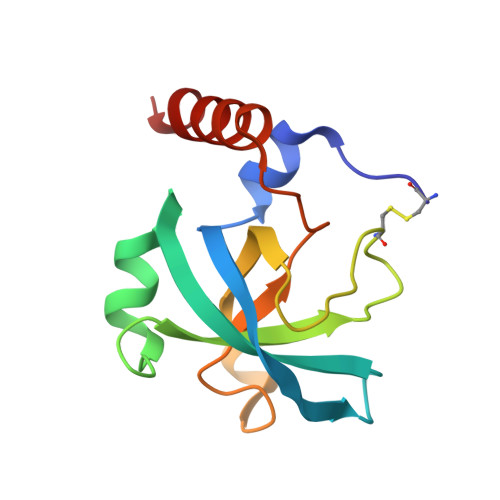
An interdomain disulfide bridge links the NtA and first FS domain in agrin.

McFarlane, A.A.Stetefeld, J.

(2009) Protein Sci 18: 2421-2428

  • DOI: https://doi.org/10.1002/pro.276
  • Primary Citation of Related Structures:  
    3I70

  • PubMed Abstract: 

    Agrin is a multidomain heparan sulfate proteoglycan involved in postsynaptic differentiation at the neuromuscular junction. Binding of agrin to synaptic basal lamina is mediated by the N-terminal agrin (NtA) domain. The NtA domain of agrin is followed by a tandem of nine follistatin-like (FS) domains forming a rod-like spacer to the laminin G-like domains of the molecule. Here we report that the most C-terminal cysteine residue of NtA (Cys123) forms an interdomain disulfide bond with the FOLN subdomain of the FS module. Remarkably, this single cysteine is flanked by Leu117 and Val124, which are two essential beta-branched amino acids forming the heterocomplex of NtA with the gamma 1 chain of laminin. Moreover, we show that this covalent linkage compensates for the seven amino acid residue splice insert at the very C-terminal helix H3 and causes a rigid interface between NtA and FS independent of the alternative mRNA splice event. These results suggest that the interdomain disulfide bond between the NtA and the first FS domain might be important for the proper folding of agrin.


  • Organizational Affiliation

    Department of Chemistry, University of Manitoba, Winnipeg, Manitoba R3T 2N2, Canada.


Macromolecules
Find similar proteins by:  (by identity cutoff)  |  3D Structure
Entity ID: 1
MoleculeChains Sequence LengthOrganismDetailsImage
Agrin128Gallus gallusMutation(s): 0 
UniProt
Find proteins for P31696 (Gallus gallus)
Explore P31696 
Go to UniProtKB:  P31696
Entity Groups  
Sequence Clusters30% Identity50% Identity70% Identity90% Identity95% Identity100% Identity
UniProt GroupP31696
Sequence Annotations
Expand
  • Reference Sequence
Experimental Data & Validation

Experimental Data

  • Method: X-RAY DIFFRACTION
  • Resolution: 2.30 Å
  • R-Value Free: 0.276 
  • R-Value Work: 0.221 
  • R-Value Observed: 0.221 
  • Space Group: C 1 2 1
Unit Cell:
Length ( Å )Angle ( ˚ )
a = 80.229α = 90
b = 50.71β = 117.09
c = 54.187γ = 90
Software Package:
Software NamePurpose
CNSrefinement

Structure Validation

View Full Validation Report


Entry History 

Deposition Data

Revision History  (Full details and data files)

  • Version 1.0: 2010-03-02
    Type: Initial release
  • Version 1.1: 2011-07-13
    Changes: Version format compliance
  • Version 1.2: 2018-04-18
    Changes: Data collection
  • Version 1.3: 2023-09-06
    Changes: Data collection, Database references, Refinement description