


Crystal structure of periplasmic binding ribose operon repressor protein from Lactobacillus acidophilus
Agarwal, R., Burley, S.K., Swaminathan, S.To be published.
Experimental Data Snapshot
wwPDB Validation 3D Report Full Report

Entity ID: 1 | |||||
|---|---|---|---|---|---|
| Molecule | Chains | Sequence Length | Organism | Details | Image |
| Ribose operon repressor | 277 | Lactobacillus acidophilus | Mutation(s): 0 Gene Names: rbsR, LBA1472 | 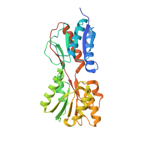 | |
UniProt | |||||
Find proteins for Q5FJ32 (Lactobacillus acidophilus (strain ATCC 700396 / NCK56 / N2 / NCFM)) Explore Q5FJ32 Go to UniProtKB: Q5FJ32 | |||||
Entity Groups | |||||
| Sequence Clusters | 30% Identity50% Identity70% Identity90% Identity95% Identity100% Identity | ||||
| UniProt Group | Q5FJ32 | ||||
Sequence AnnotationsExpand | |||||
| |||||
| Length ( Å ) | Angle ( ˚ ) |
|---|---|
| a = 59.597 | α = 90 |
| b = 70.224 | β = 90 |
| c = 139.439 | γ = 90 |
| Software Name | Purpose |
|---|---|
| CBASS | data collection |
| SHELXD | phasing |
| SHARP | phasing |
| CNS | refinement |
| HKL-2000 | data reduction |
| HKL-2000 | data scaling |
| SCALEPACK | data scaling |
RCSB PDB (citation) is hosted by
RCSB PDB is a member of the

