Molecular recognition of physiological substrate noradrenaline by the adrenaline-synthesizing enzyme PNMT and factors influencing its methyltransferase activity.
Drinkwater, N., Gee, C.L., Puri, M., Criscione, K.R., McLeish, M.J., Grunewald, G.L., Martin, J.L.(2009) Biochem J 422: 463-471
- PubMed: 19570037
- DOI: https://doi.org/10.1042/BJ20090702
- Primary Citation of Related Structures:
3HCA, 3HCB, 3HCC, 3HCD, 3HCE, 3HCF - PubMed Abstract:
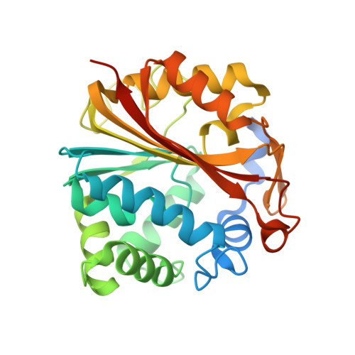
Substrate specificity is critically important for enzyme catalysis. In the adrenaline-synthesizing enzyme PNMT (phenylethanolamine N-methyltransferase), minor changes in substituents can convert substrates into inhibitors. Here we report the crystal structures of six human PNMT complexes, including the first structure of the enzyme in complex with its physiological ligand R-noradrenaline. Determining this structure required rapid soak methods because of the tendency for noradrenaline to oxidize. Comparison of the PNMT-noradrenaline complex with the previously determined PNMT-p-octopamine complex demonstrates that these two substrates form almost equivalent interactions with the enzyme and show that p-octopamine is a valid model substrate for PNMT. The crystal structures illustrate the adaptability of the PNMT substrate binding site in accepting multi-fused ring systems, such as substituted norbornene, as well as noradrenochrome, the oxidation product of noradrenaline. These results explain why only a subset of ligands recognized by PNMT are methylated by the enzyme; bulky substituents dictate the binding orientation of the ligand and can thereby place the acceptor amine too far from the donor methyl group for methylation to occur. We also show how the critical Glu(185) catalytic residue can be replaced by aspartic acid with a loss of only 10-fold in catalytic efficiency. This is because protein backbone movements place the Asp(185) carboxylate almost coincident with the carboxylate of Glu(185). Conversely, replacement of Glu(185) by glutamine reduces catalytic efficiency almost 300-fold, not only because of the loss of charge, but also because the variant residue does not adopt the same conformation as Glu(185).
Organizational Affiliation:
Institute for Molecular Bioscience, Division of Chemistry and Structural Biology, The University of Queensland, Brisbane, Queensland 4072, Australia.