Biochemical and structural characterization of alanine racemase from Bacillus anthracis (Ames).
Counago, R.M., Davlieva, M., Strych, U., Hill, R.E., Krause, K.L.(2009) BMC Struct Biol 9: 53-53
- PubMed: 19695097
- DOI: https://doi.org/10.1186/1472-6807-9-53
- Primary Citation of Related Structures:
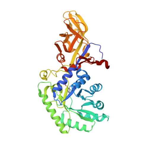
3HA1 - PubMed Abstract:
Bacillus anthracis is the causative agent of anthrax and a potential bioterrorism threat. Here we report the biochemical and structural characterization of B. anthracis (Ames) alanine racemase (AlrBax), an essential enzyme in prokaryotes and a target for antimicrobial drug development. We also compare the native AlrBax structure to a recently reported structure of the same enzyme obtained through reductive lysine methylation. B. anthracis has two open reading frames encoding for putative alanine racemases. We show that only one, dal1, is able to complement a D-alanine auxotrophic strain of E. coli. Purified Dal1, which we term AlrBax, is shown to be a dimer in solution by dynamic light scattering and has a Vmax for racemization (L- to D-alanine) of 101 U/mg. The crystal structure of unmodified AlrBax is reported here to 1.95 A resolution. Despite the overall similarity of the fold to other alanine racemases, AlrBax makes use of a chloride ion to position key active site residues for catalysis, a feature not yet observed for this enzyme in other species. Crystal contacts are more extensive in the methylated structure compared to the unmethylated structure. The chloride ion in AlrBax is functioning effectively as a carbamylated lysine making it an integral and unique part of this structure. Despite differences in space group and crystal form, the two AlrBax structures are very similar, supporting the case that reductive methylation is a valid rescue strategy for proteins recalcitrant to crystallization, and does not, in this case, result in artifacts in the tertiary structure.
Organizational Affiliation:
Department of Biochemistry, University of Otago, Dunedin, New Zealand. rafael.counago@otago.ac.nz