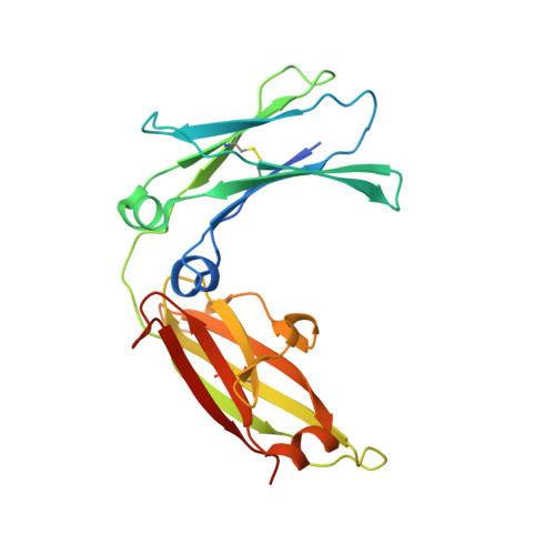
POP-OUT | CLOSE
Crystal structure of the IgE-Fc3-4 domains
3H9Y
Primary Citation
 
 
  •   Related Citations in PDB Entry (REMARK 1) Hide
     
  •   Molecular Description Hide
    Classification: Immune System
    Structure Weight: 77673.57
    Molecule: Ig epsilon chain C region
    Polymer: 1 Type: protein Length: 223
    Chains: A, B, E
    Fragment: UNP residues 209-428
    Mutation: N252Q, N264Q
    Organism Homo sapiens
    Gene Name IGHE
    UniProtKB:   Protein Feature View | Search PDB | P01854  
     
  •   Source Hide
    Polymer: 1
    Scientific Name: Homo sapiens   Taxonomy   Common Name: Human Expression System: Trichoplusia ni  
    Polymer: 2
    Scientific Name: Synthetic construct   Taxonomy    
    Polymer: 3
    Scientific Name: Synthetic construct   Taxonomy    
     
  •   Related PDB Entries Hide
    Identifier Details
    1F6A  The 'wild-type carbohydrate' form of the same protein in complex with the alpha chain of the IgE-FcRI. 
    1FP5  The 'wild-type carbohydrate' form of the same protein (has wild-type N371 and N383). 
    1O0V  The structure of the IgE-Fc domains 2-4. 
    3H9Z  Crystal structure of the IgE-Fc3-4 domains, crystal form 2 
    3HA0  Crystal structure of the IgE-Fc3-4 domains, crystal form 3 
     
  •   Ligand Chemical Component Hide
    Identifier Formula Name Interactions
    BMA
    Search 
    Download 
    BMA C6 H12 O6
    BETA-D-MANNOSE
    MAN
    Search 
    Download 
    MAN C6 H12 O6
    ALPHA-D-MANNOSE
    NAG
    Search 
    Download 
    NAG C8 H15 N O6
    N-ACETYL-D-GLUCOSAMINE
    NH4
    Search 
    Download 
    NH4 H4 N
    AMMONIUM ION
     
  •   External Domain Annotations Hide
     
  •   Structural Biology Knowledgebase Data Hide
     
 
Data in orange boxes are gathered from external resources (when available).
 
  Biological Assembly 1       
Biological assembly 1 assigned by authors and generated by PISA (software)
Downloadable viewers:


  •   MyPDB Personal Annotations Hide
     
  •   Deposition Summary Hide
    Authors:   Wurzburg, B.A.

    Deposition:   2009-04-30
    Release:   2009-09-08
    Last Modified (REVDAT):   2011-07-13
     
  •   Revision History    Hide
    Mouse over text for details
    2011-07-13
    Flag residual B-value
    2011-07-13
    Refinement description
    2011-07-13
    Version format compliance
     
  •   Experimental Details Hide
    Method:   X-RAY DIFFRACTION
    Exp. Data:
      Structure Factors
    EDS  
    Resolution[Å]:   2.23
    R-Value: 0.233 (obs.)
    R-Free: 0.262
    Space Group: C 2
    Unit Cell:
      Length [Å] Angles [°]
    a = 153.68 α = 90.00 
    b = 104.99 β = 101.61 
    c = 49.22 γ = 90.00