3GD9

Crystal structure of laminaripentaose-producing beta-1,3-glucanase in complex with laminaritetraose


Experimental Data Snapshot

  • Method: X-RAY DIFFRACTION
  • Resolution: 1.80 Å
  • R-Value Free: 0.217 
  • R-Value Work: 0.166 
  • R-Value Observed: 0.172 

wwPDB Validation   3D Report Full Report


This is version 2.1 of the entry. See complete history


Literature

Structure, mechanistic action, and essential residues of a GH-64 enzyme, laminaripentaose-producing beta-1,3-glucanase.

Wu, H.M.Liu, S.W.Hsu, M.T.Hung, C.L.Lai, C.C.Cheng, W.C.Wang, H.J.Li, Y.K.Wang, W.C.

(2009) J Biol Chem 284: 26708-26715

  • DOI: https://doi.org/10.1074/jbc.M109.010983
  • Primary Citation of Related Structures:  
    3GD0, 3GD9

  • PubMed Abstract: 

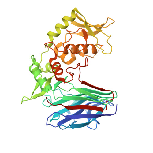
    Laminaripentaose-producing beta-1,3-glucanase (LPHase), a member of glycoside hydrolase family 64, cleaves a long-chain polysaccharide beta-1,3-glucan into specific pentasaccharide oligomers. The crystal structure of LPHase from Streptomyces matensis DIC-108 was solved to 1.62 A resolution using multiple-wavelength anomalous dispersion methods. The LPHase structure reveals a novel crescent-like fold; it consists of a barrel domain and a mixed (alpha/beta) domain, forming a wide-open groove between the two domains. The liganded crystal structure was also solved to 1.80 A, showing limited conformational changes. Within the wide groove, a laminaritetraose molecule is found to sit in an electronegatively charged central region and is proximal to several conserved residues including two carboxylates (Glu(154) and Asp(170)) and four other sugar-binding residues (Thr(156), Asn(158), Trp(163), and Thr(167)). Molecular modeling using a laminarihexaose as a substrate suggests roles for Glu(154) and Asp(170) as acid and base catalysts, respectively, whereas the side chains of Thr(156), Asn(158), and Trp(163) demarcate subsite +5. Site-directed mutagenesis of Glu(154) and Asp(170) confirms that both carboxylates are essential for catalysis. Together, our results suggest that LPHase uses a direct displacement mechanism involving Glu(154) and Asp(170) to cleave a beta-1,3-glucan into specific alpha-pentasaccharide oligomers.


  • Organizational Affiliation

    Department of Life Science, Institute of Molecular and Cellular Biology, National Tsing Hua University, Hsinchu 300, Taiwan.


Macromolecules
Find similar proteins by:  (by identity cutoff)  |  3D Structure
Entity ID: 1
MoleculeChains Sequence LengthOrganismDetailsImage
Laminaripentaose-producing beta-1,3-guluase (LPHase)367Streptomyces matensisMutation(s): 0 
Gene Names: LPHase
EC: 3.2.1.39
UniProt
Find proteins for Q9Z4I2 (Streptomyces matensis)
Explore Q9Z4I2 
Go to UniProtKB:  Q9Z4I2
Entity Groups  
Sequence Clusters30% Identity50% Identity70% Identity90% Identity95% Identity100% Identity
UniProt GroupQ9Z4I2
Sequence Annotations
Expand
  • Reference Sequence
Oligosaccharides

Help

Entity ID: 2
MoleculeChains Length2D Diagram Glycosylation3D Interactions
beta-D-glucopyranose-(1-3)-beta-D-glucopyranose-(1-3)-beta-D-glucopyranose-(1-3)-alpha-D-glucopyranose
B
4N/A
Glycosylation Resources
GlyTouCan:  G38015EF
GlyCosmos:  G38015EF
GlyGen:  G38015EF
Experimental Data & Validation

Experimental Data

  • Method: X-RAY DIFFRACTION
  • Resolution: 1.80 Å
  • R-Value Free: 0.217 
  • R-Value Work: 0.166 
  • R-Value Observed: 0.172 
  • Space Group: P 21 21 21
Unit Cell:
Length ( Å )Angle ( ˚ )
a = 45.981α = 90
b = 60.352β = 90
c = 149.118γ = 90
Software Package:
Software NamePurpose
HKL-2000data collection
MOLREPphasing
REFMACrefinement
HKL-2000data reduction
HKL-2000data scaling

Structure Validation

View Full Validation Report


Entry History 

Deposition Data

Revision History  (Full details and data files)

  • Version 1.0: 2009-07-28
    Type: Initial release
  • Version 1.1: 2011-07-13
    Changes: Version format compliance
  • Version 2.0: 2020-07-29
    Type: Remediation
    Reason: Carbohydrate remediation
    Changes: Advisory, Atomic model, Data collection, Database references, Derived calculations, Structure summary
  • Version 2.1: 2023-11-01
    Changes: Data collection, Database references, Refinement description, Structure summary