3F9P

Crystal structure of myeloperoxidase from human leukocytes


Experimental Data Snapshot

  • Method: X-RAY DIFFRACTION
  • Resolution: 2.93 Å
  • R-Value Free: 0.257 
  • R-Value Work: 0.236 
  • R-Value Observed: 0.237 

wwPDB Validation   3D Report Full Report


Ligand Structure Quality Assessment 


This is version 2.1 of the entry. See complete history


Literature

Essential role of proximal histidine-asparagine interaction in Mammalian peroxidases.

Carpena, X.Vidossich, P.Schroettner, K.Calisto, B.M.Banerjee, S.Stampler, J.Soudi, M.Furtmuller, P.G.Rovira, C.Fita, I.Obinger, C.

(2009) J Biol Chem 284: 25929-25937

  • DOI: https://doi.org/10.1074/jbc.M109.002154
  • Primary Citation of Related Structures:  
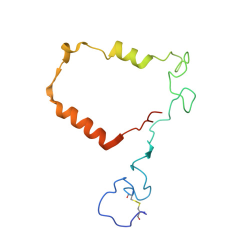
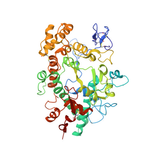
    3F9P

  • PubMed Abstract: 

    In heme enzymes belonging to the peroxidase-cyclooxygenase superfamily the proximal histidine is in close interaction with a fully conserved asparagine. The crystal structure of a mixture of glycoforms of myeloperoxidase (MPO) purified from granules of human leukocytes prompted us to revise the orientation of this asparagine and the protonation status of the proximal histidine. The data we present contrast with previous MPO structures, but are strongly supported by molecular dynamics simulations. Moreover, comprehensive analysis of published lactoperoxidase structures suggest that the described proximal heme architecture is a general structural feature of animal heme peroxidases. Its importance is underlined by the fact that the MPO variant N421D, recombinantly expressed in mammalian cell lines, exhibited modified spectral properties and diminished catalytic activity compared with wild-type recombinant MPO. It completely lost its ability to oxidize chloride to hypochlorous acid, which is a characteristic feature of MPO and essential for its role in host defense. The presented crystal structure of MPO revealed further important differences compared with the published structures including the extent of glycosylation, interaction between light and heavy polypeptides, as well as heme to protein covalent bonds. These data are discussed with respect to biosynthesis and post-translational maturation of MPO as well as to its peculiar biochemical and biophysical properties.


  • Organizational Affiliation

    Institute of Research in Biomedicine (IRB-Barcelona), Parc Científic, Baldiri i Reixac 10, 08028 Barcelona, Spain.


Macromolecules
Find similar proteins by:  (by identity cutoff)  |  3D Structure
Entity ID: 1
MoleculeChains Sequence LengthOrganismDetailsImage
MyeloperoxidaseA,
C [auth B]
114Homo sapiensMutation(s): 0 
EC: 1.11.1.7
UniProt & NIH Common Fund Data Resources
Find proteins for P05164 (Homo sapiens)
Explore P05164 
Go to UniProtKB:  P05164
PHAROS:  P05164
GTEx:  ENSG00000005381 
Entity Groups  
Sequence Clusters30% Identity50% Identity70% Identity90% Identity95% Identity100% Identity
UniProt GroupP05164
Sequence Annotations
Expand
  • Reference Sequence
Find similar proteins by:  (by identity cutoff)  |  3D Structure
Entity ID: 2
MoleculeChains Sequence LengthOrganismDetailsImage
MyeloperoxidaseB [auth C],
D
467Homo sapiensMutation(s): 0 
EC: 1.11.1.7
UniProt & NIH Common Fund Data Resources
Find proteins for P05164 (Homo sapiens)
Explore P05164 
Go to UniProtKB:  P05164
PHAROS:  P05164
GTEx:  ENSG00000005381 
Entity Groups  
Sequence Clusters30% Identity50% Identity70% Identity90% Identity95% Identity100% Identity
UniProt GroupP05164
Sequence Annotations
Expand
  • Reference Sequence
Oligosaccharides

Help

Entity ID: 3
MoleculeChains Length2D Diagram Glycosylation3D Interactions
beta-D-mannopyranose-(1-3)-[beta-D-mannopyranose-(1-6)]beta-D-mannopyranose-(1-4)-2-acetamido-2-deoxy-beta-D-glucopyranose-(1-4)-[alpha-L-fucopyranose-(1-6)]2-acetamido-2-deoxy-beta-D-glucopyranose
E, F
6N-Glycosylation
Glycosylation Resources
GlyTouCan:  G38085DZ
GlyCosmos:  G38085DZ
GlyGen:  G38085DZ
Small Molecules
Ligands 5 Unique
IDChains Name / Formula / InChI Key2D Diagram3D Interactions
HEM
Query on HEM

Download Ideal Coordinates CCD File 
H [auth A],
M [auth B]
PROTOPORPHYRIN IX CONTAINING FE
C34 H32 Fe N4 O4
KABFMIBPWCXCRK-RGGAHWMASA-L
NAG
Query on NAG

Download Ideal Coordinates CCD File 
J [auth C],
K [auth C],
P [auth D],
Q [auth D]
2-acetamido-2-deoxy-beta-D-glucopyranose
C8 H15 N O6
OVRNDRQMDRJTHS-FMDGEEDCSA-N
ACT
Query on ACT

Download Ideal Coordinates CCD File 
L [auth C],
R [auth D]
ACETATE ION
C2 H3 O2
QTBSBXVTEAMEQO-UHFFFAOYSA-M
CA
Query on CA

Download Ideal Coordinates CCD File 
I [auth C],
N [auth D]
CALCIUM ION
Ca
BHPQYMZQTOCNFJ-UHFFFAOYSA-N
CL
Query on CL

Download Ideal Coordinates CCD File 
G [auth A],
O [auth D]
CHLORIDE ION
Cl
VEXZGXHMUGYJMC-UHFFFAOYSA-M
Modified Residues  1 Unique
IDChains TypeFormula2D DiagramParent
CSO
Query on CSO
B [auth C],
D
L-PEPTIDE LINKINGC3 H7 N O3 SCYS
Experimental Data & Validation

Experimental Data

  • Method: X-RAY DIFFRACTION
  • Resolution: 2.93 Å
  • R-Value Free: 0.257 
  • R-Value Work: 0.236 
  • R-Value Observed: 0.237 
  • Space Group: P 43 21 2
Unit Cell:
Length ( Å )Angle ( ˚ )
a = 110.74α = 90
b = 110.74β = 90
c = 255.333γ = 90
Software Package:
Software NamePurpose
ADSCdata collection
MOLREPphasing
REFMACrefinement
SCALEPACKdata scaling
DENZOdata reduction

Structure Validation

View Full Validation Report


Ligand Structure Quality Assessment 


Entry History 

Deposition Data

Revision History  (Full details and data files)

  • Version 1.0: 2009-07-14
    Type: Initial release
  • Version 1.1: 2011-07-13
    Changes: Non-polymer description, Version format compliance
  • Version 2.0: 2020-07-29
    Type: Remediation
    Reason: Carbohydrate remediation
    Changes: Atomic model, Data collection, Derived calculations, Structure summary
  • Version 2.1: 2023-12-27
    Changes: Data collection, Database references, Structure summary