3F2M

Urate oxidase complexed with 8-azaxanthine at 150 MPa


Experimental Data Snapshot

  • Method: X-RAY DIFFRACTION
  • Resolution: 1.80 Å
  • R-Value Free: 0.218 
  • R-Value Work: 0.182 
  • R-Value Observed: 0.184 

wwPDB Validation   3D Report Full Report


Ligand Structure Quality Assessment 


This is version 1.4 of the entry. See complete history


Literature

Structure-function perturbation and dissociation of tetrameric urate oxidase by high hydrostatic pressure.

Girard, E.Marchal, S.Perez, J.Finet, S.Kahn, R.Fourme, R.Marassio, G.Dhaussy, A.C.Prange, T.Giffard, M.Dulin, F.Bonnete, F.Lange, R.Abraini, J.H.Mezouar, M.Colloc'h, N.

(2010) Biophys J 98: 2365-2373

  • DOI: https://doi.org/10.1016/j.bpj.2010.01.058
  • Primary Citation of Related Structures:  
    3F2M

  • PubMed Abstract: 

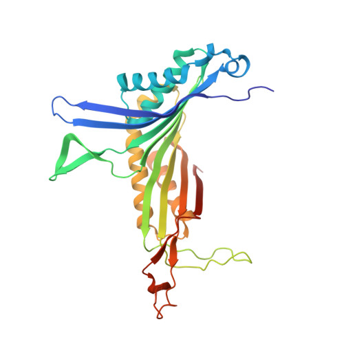
    Structure-function relationships in the tetrameric enzyme urate oxidase were investigated using pressure perturbation. As the active sites are located at the interfaces between monomers, enzyme activity is directly related to the integrity of the tetramer. The effect of hydrostatic pressure on the enzyme was investigated by x-ray crystallography, small-angle x-ray scattering, and fluorescence spectroscopy. Enzymatic activity was also measured under pressure and after decompression. A global model, consistent with all measurements, discloses structural and functional details of the pressure-induced dissociation of the tetramer. Before dissociating, the pressurized protein adopts a conformational substate characterized by an expansion of its substrate binding pocket at the expense of a large neighboring hydrophobic cavity. This substate should be adopted by the enzyme during its catalytic mechanism, where the active site has to accommodate larger intermediates and product. The approach, combining several high-pressure techniques, offers a new (to our knowledge) means of exploring structural and functional properties of transient states relevant to protein mechanisms.


  • Organizational Affiliation

    Institut de Biologie Structurale J.-P. Ebel UMR 5075 CEA CNRS UJF, Grenoble, France.


Macromolecules
Find similar proteins by:  (by identity cutoff)  |  3D Structure
Entity ID: 1
MoleculeChains Sequence LengthOrganismDetailsImage
Uricase302Aspergillus flavusMutation(s): 1 
Gene Names: uaZuox
EC: 1.7.3.3
UniProt
Find proteins for Q00511 (Aspergillus flavus)
Explore Q00511 
Go to UniProtKB:  Q00511
Entity Groups  
Sequence Clusters30% Identity50% Identity70% Identity90% Identity95% Identity100% Identity
UniProt GroupQ00511
Sequence Annotations
Expand
  • Reference Sequence
Small Molecules
Experimental Data & Validation

Experimental Data

  • Method: X-RAY DIFFRACTION
  • Resolution: 1.80 Å
  • R-Value Free: 0.218 
  • R-Value Work: 0.182 
  • R-Value Observed: 0.184 
  • Space Group: I 2 2 2
Unit Cell:
Length ( Å )Angle ( ˚ )
a = 79.7α = 90
b = 95.87β = 90
c = 104.99γ = 90
Software Package:
Software NamePurpose
SCALAdata scaling
REFMACrefinement
PDB_EXTRACTdata extraction
specdata collection
XDSdata reduction
REFMACphasing

Structure Validation

View Full Validation Report


Ligand Structure Quality Assessment 


Entry History 

Deposition Data

Revision History  (Full details and data files)

  • Version 1.0: 2009-11-10
    Type: Initial release
  • Version 1.1: 2011-07-13
    Changes: Version format compliance
  • Version 1.2: 2014-02-19
    Changes: Database references
  • Version 1.3: 2021-09-15
    Changes: Database references, Derived calculations, Structure summary
  • Version 1.4: 2023-11-01
    Changes: Data collection, Refinement description