3EVS

Crystal structure of the GDF-5:BMP receptor IB complex.


Experimental Data Snapshot

  • Method: X-RAY DIFFRACTION
  • Resolution: 2.10 Å
  • R-Value Free: 0.256 
  • R-Value Work: 0.215 
  • R-Value Observed: 0.217 

wwPDB Validation   3D Report Full Report


This is version 1.2 of the entry. See complete history


Literature

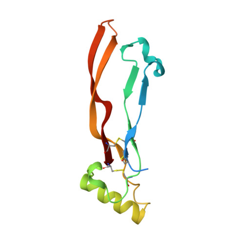
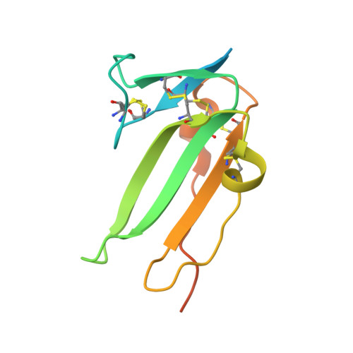
Crystal structure analysis reveals a spring-loaded latch as molecular mechanism for GDF-5-type I receptor specificity.

Kotzsch, A.Nickel, J.Seher, A.Sebald, W.Muller, T.D.

(2009) EMBO J 28: 937-947

  • DOI: https://doi.org/10.1038/emboj.2009.37
  • Primary Citation of Related Structures:  
    3EVS

  • PubMed Abstract: 

    Dysregulation of growth and differentiation factor 5 (GDF-5) signalling, a member of the TGF-beta superfamily, is strongly linked to skeletal malformation. GDF-5-mediated signal transduction involves both BMP type I receptors, BMPR-IA and BMPR-IB. However, mutations in either GDF-5 or BMPR-IB lead to similar phenotypes, indicating that in chondrogenesis GDF-5 signalling seems to be exclusively mediated through BMPR-IB. Here, we present structural insights into the GDF-5:BMPR-IB complex revealing how binding specificity for BMPR-IB is generated on a molecular level. In BMPR-IB, a loop within the ligand-binding epitope functions similar to a latch allowing high-affinity binding of GDF-5. In BMPR-IA, this latch is in a closed conformation leading to steric repulsion. The new structural data now provide also a molecular basis of how phenotypically relevant missense mutations in GDF-5 might impair receptor binding and activation.


  • Organizational Affiliation

    Lehrstuhl für Botanik I-Molekulare Pflanzenphysiologie und Biophysik, Julius-von-Sachs-Institut für Biowissenschaften (Biozentrum) der Universität Würzburg, Würzburg, Germany.


Macromolecules
Find similar proteins by:  (by identity cutoff)  |  3D Structure
Entity ID: 1
MoleculeChains Sequence LengthOrganismDetailsImage
Growth/differentiation factor 5A [auth B]117Homo sapiensMutation(s): 0 
Gene Names: CDMP1GDF5
UniProt & NIH Common Fund Data Resources
Find proteins for P43026 (Homo sapiens)
Explore P43026 
Go to UniProtKB:  P43026
PHAROS:  P43026
GTEx:  ENSG00000125965 
Entity Groups  
Sequence Clusters30% Identity50% Identity70% Identity90% Identity95% Identity100% Identity
UniProt GroupP43026
Sequence Annotations
Expand
  • Reference Sequence
Find similar proteins by:  (by identity cutoff)  |  3D Structure
Entity ID: 2
MoleculeChains Sequence LengthOrganismDetailsImage
Bone morphogenetic protein receptor type-1BB [auth C]119Mus musculusMutation(s): 0 
Gene Names: Acvrlk6Bmpr1b
UniProt & NIH Common Fund Data Resources
Find proteins for P36898 (Mus musculus)
Explore P36898 
Go to UniProtKB:  P36898
IMPC:  MGI:107191
Entity Groups  
Sequence Clusters30% Identity50% Identity70% Identity90% Identity95% Identity100% Identity
UniProt GroupP36898
Sequence Annotations
Expand
  • Reference Sequence
Experimental Data & Validation

Experimental Data

  • Method: X-RAY DIFFRACTION
  • Resolution: 2.10 Å
  • R-Value Free: 0.256 
  • R-Value Work: 0.215 
  • R-Value Observed: 0.217 
  • Space Group: P 42 21 2
Unit Cell:
Length ( Å )Angle ( ˚ )
a = 76.455α = 90
b = 76.455β = 90
c = 82.784γ = 90
Software Package:
Software NamePurpose
d*TREKdata scaling
SHARPphasing
DMphasing
REFMACrefinement
PDB_EXTRACTdata extraction
MAR345dtbdata collection
CrystalCleardata reduction
CrystalCleardata scaling

Structure Validation

View Full Validation Report


Entry History 

Deposition Data

Revision History  (Full details and data files)

  • Version 1.0: 2009-03-10
    Type: Initial release
  • Version 1.1: 2011-07-13
    Changes: Advisory, Version format compliance
  • Version 1.2: 2023-12-27
    Changes: Data collection, Database references