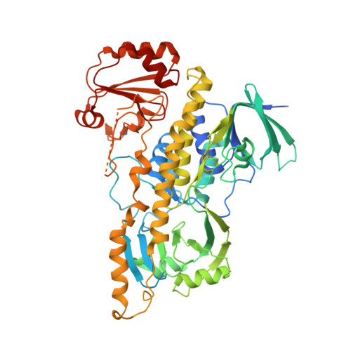
The FAD cofactor of RebC shifts to an IN conformation upon flavin reduction
Ryan, K.S., Chakraborty, S., Howard-Jones, A.R., Walsh, C.T., Ballou, D.P., Drennan, C.L.(2008) Biochemistry 47: 13506-13513
- PubMed: 19035832
- DOI: https://doi.org/10.1021/bi801229w
- Primary Citation of Related Structures:
3EPT - PubMed Abstract:
RebC is a putative flavin hydroxylase functioning together with RebP to carry out a key step in the biosynthesis of rebeccamycin. To probe the mechanism of flavin-based chemistry in RebC, we solved the structure of RebC with reduced flavin. Upon flavin reduction, the RebC crystal undergoes a change in its unit cell dimension concurrent with a 5 A movement of the isoalloxazine ring, positioning the flavin ring adjacent to the substrate-binding pocket. Additionally, a disordered helix becomes ordered upon flavin reduction, closing off one side of the substrate-binding pocket. This structure, along with previously reported structures, increases our understanding of the RebC enzyme mechanism, indicating that either the reduction of the flavin itself or binding of substrate is sufficient to drive major conformational changes in RebC to generate a closed active site. Our finding that flavin reduction seals the active site such that substrate cannot enter suggests that our reduced flavin RebC structure is off-pathway and that substrate binding is likely to precede flavin reduction during catalysis. Along with kinetic data presented here, these structures suggest that the first cycle of catalysis in RebC may resemble that of p-hydroxybenzoate hydroxylase, with substrate binding promoting flavin reduction.
Organizational Affiliation:
Department of Biology, Massachusetts Institute of Technology, Cambridge, Massachusetts 02139, USA.