3ENZ

Arsenolytic structure of Plasmodium falciparum purine nucleoside phosphorylase with hypoxanthine, ribose and arsenate ion


Experimental Data Snapshot

  • Method: X-RAY DIFFRACTION
  • Resolution: 2.03 Å
  • R-Value Free: 0.193 
  • R-Value Work: 0.160 
  • R-Value Observed: 0.160 

wwPDB Validation   3D Report Full Report


This is version 2.1 of the entry. See complete history


Literature

Conservation of structure and activity in Plasmodium purine nucleoside phosphorylases.

Chaikuad, A.Brady, R.L.

(2009) BMC Struct Biol 9: 42-42

  • DOI: https://doi.org/10.1186/1472-6807-9-42
  • Primary Citation of Related Structures:  
    3EMV, 3ENZ

  • PubMed Abstract: 

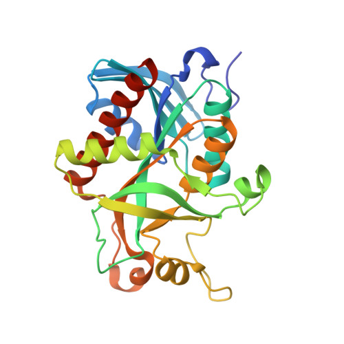
    Purine nucleoside phosphorylase (PNP) is central to purine salvage mechanisms in Plasmodium parasites, the causative agents of malaria. Most human malaria results from infection either by Plasmodium falciparum (Pf), the deadliest form of the parasite, or by the widespread Plasmodium vivax (Pv). Whereas the PNP enzyme from Pf has previously been studied in detail, despite the prevalence of Pv little is known about many of the key metabolic enzymes from this parasite, including PvPNP. The crystal structure of PvPNP is described and is seen to have many features in common with the previously reported structure of PfPNP. In particular, the composition and conformations of the active site regions are virtually identical. The crystal structure of a complex of PfPNP co-crystallised with inosine and arsenate is also described, and is found to contain a mixture of products and reactants - hypoxanthine, ribose and arsenate. The ribose C1' in this hybrid complex lies close to the expected point of symmetry along the PNP reaction coordinate, consistent with a conformation between the transition and product states. These two Plasmodium PNP structures confirm the similarity of structure and mechanism of these enzymes, which are also confirmed in enzyme kinetic assays using an array of substrates. These reveal an unusual form of substrate activation by 2'-deoxyinosine of PvPNP, but not PfPNP. The close similarity of the Pf and Pv PNP structures allows characteristic features to be identified that differentiate the Apicomplexa PNPs from the human host enzyme. This similarity also suggests there should be a high level of cross-reactivity for compounds designed to inhibit either of these molecular targets. However, despite these similarities, there are also small differences in the activities of the two Plasmodium enzymes.


  • Organizational Affiliation

    Department of Biochemistry, University of Bristol, Bristol, BS8 1TD, UK. Apirat.Chaikuad@sgc.ox.ac.uk


Macromolecules
Find similar proteins by:  (by identity cutoff)  |  3D Structure
Entity ID: 1
MoleculeChains Sequence LengthOrganismDetailsImage
Purine nucleoside phosphorylase
A, B, C, D, E
A, B, C, D, E, F
253Plasmodium falciparum 3D7Mutation(s): 0 
Gene Names: PFE0660c
EC: 2.4.2.1
UniProt
Find proteins for Q8I3X4 (Plasmodium falciparum (isolate 3D7))
Explore Q8I3X4 
Go to UniProtKB:  Q8I3X4
Entity Groups  
Sequence Clusters30% Identity50% Identity70% Identity90% Identity95% Identity100% Identity
UniProt GroupQ8I3X4
Sequence Annotations
Expand
  • Reference Sequence
Small Molecules
Ligands 5 Unique
IDChains Name / Formula / InChI Key2D Diagram3D Interactions
ART
Query on ART

Download Ideal Coordinates CCD File 
I [auth A]
JA [auth D]
T [auth B]
TA [auth E]
XA [auth F]
I [auth A],
JA [auth D],
T [auth B],
TA [auth E],
XA [auth F],
Z [auth C]
ARSENATE
As O4
DJHGAFSJWGLOIV-UHFFFAOYSA-K
HPA
Query on HPA

Download Ideal Coordinates CCD File 
G [auth A]
HA [auth D]
R [auth B]
RA [auth E]
VA [auth F]
G [auth A],
HA [auth D],
R [auth B],
RA [auth E],
VA [auth F],
X [auth C]
HYPOXANTHINE
C5 H4 N4 O
FDGQSTZJBFJUBT-UHFFFAOYSA-N
R1X
Query on R1X

Download Ideal Coordinates CCD File 
H [auth A]
IA [auth D]
S [auth B]
SA [auth E]
WA [auth F]
H [auth A],
IA [auth D],
S [auth B],
SA [auth E],
WA [auth F],
Y [auth C]
1,4-anhydro-D-ribitol
C5 H10 O4
KZVAAIRBJJYZOW-LMVFSUKVSA-N
FMT
Query on FMT

Download Ideal Coordinates CCD File 
AA [auth C]
AB [auth F]
BA [auth C]
CA [auth C]
DA [auth C]
AA [auth C],
AB [auth F],
BA [auth C],
CA [auth C],
DA [auth C],
EA [auth C],
FA [auth C],
J [auth A],
K [auth A],
KA [auth D],
L [auth A],
LA [auth D],
M [auth A],
MA [auth D],
N [auth A],
NA [auth D],
O [auth A],
OA [auth D],
P [auth A],
U [auth B],
UA [auth E],
V [auth B],
YA [auth F],
ZA [auth F]
FORMIC ACID
C H2 O2
BDAGIHXWWSANSR-UHFFFAOYSA-N
NA
Query on NA

Download Ideal Coordinates CCD File 
GA [auth C],
PA [auth D],
Q [auth A],
QA [auth D],
W [auth B]
SODIUM ION
Na
FKNQFGJONOIPTF-UHFFFAOYSA-N
Experimental Data & Validation

Experimental Data

  • Method: X-RAY DIFFRACTION
  • Resolution: 2.03 Å
  • R-Value Free: 0.193 
  • R-Value Work: 0.160 
  • R-Value Observed: 0.160 
  • Space Group: I 41 2 2
Unit Cell:
Length ( Å )Angle ( ˚ )
a = 177.82α = 90
b = 177.82β = 90
c = 253.868γ = 90
Software Package:
Software NamePurpose
MAR345data collection
PHASERphasing
REFMACrefinement
HKL-2000data reduction
HKL-2000data scaling

Structure Validation

View Full Validation Report


Entry History 

Deposition Data

Revision History  (Full details and data files)

  • Version 1.0: 2009-08-04
    Type: Initial release
  • Version 1.1: 2011-07-13
    Changes: Advisory, Source and taxonomy, Version format compliance
  • Version 1.2: 2017-10-25
    Changes: Refinement description
  • Version 2.0: 2020-07-29
    Type: Remediation
    Reason: Carbohydrate remediation
    Changes: Advisory, Atomic model, Data collection, Database references, Derived calculations
  • Version 2.1: 2023-09-06
    Changes: Data collection, Database references, Refinement description