3DNA

Iodobenzene binding in the hydrophobic cavity of T4 lysozyme L99A mutant (seleno version)


Experimental Data Snapshot

  • Method: X-RAY DIFFRACTION
  • Resolution: 1.70 Å
  • R-Value Free: 0.217 
  • R-Value Work: 0.183 
  • R-Value Observed: 0.185 

wwPDB Validation   3D Report Full Report


Ligand Structure Quality Assessment 


This is version 1.5 of the entry. See complete history


Literature

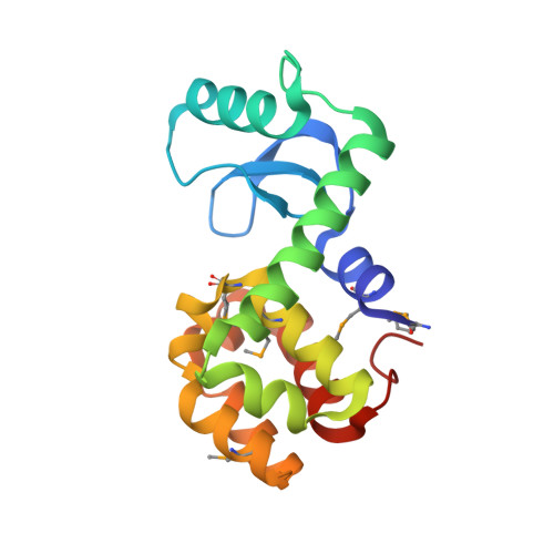
Halogenated benzenes bound within a non-polar cavity in T4 lysozyme provide examples of I...S and I...Se halogen-bonding.

Liu, L.Baase, W.A.Matthews, B.W.

(2009) J Mol Biol 385: 595-605

  • DOI: https://doi.org/10.1016/j.jmb.2008.10.086
  • Primary Citation of Related Structures:  
    3DMV, 3DMX, 3DMZ, 3DN0, 3DN1, 3DN2, 3DN3, 3DN4, 3DN6, 3DN8, 3DNA

  • PubMed Abstract: 

    We showed earlier that the mutation of Leu99 to alanine in bacteriophage T4 lysozyme creates an internal cavity of volume approximately 150 A(3) that binds benzene and a variety of other ligands. As such, this cavity provides an excellent target to study protein-ligand interaction. Here, we use low-temperature crystallography and related techniques to analyze the binding of halogen-incorporated benzenes typified by C(6)F(5)X, where X=H, F, Cl, Br or I, and C(6)H(5)X, where X=H or I was also studied. Because of the increased electron density of fluorine relative to hydrogen, the geometry of binding of the fluoro compounds can often be determined more precisely than their hydrogen-containing analogs. All of the ligands bind in essentially the same plane but the center of the phenyl ring can translate by up to 1.2 A. In no case does the ligand rotate freely within the cavity. The walls of the cavity consist predominantly of hydrocarbon atoms, and in several cases it appears that van der Waals interactions define the geometry of binding. In comparing the smallest with the largest ligand, the cavity volume increases from 181 A(3) to 245 A(3). This shows that the protein is flexible and adapts to the size and shape of the ligand. There is a remarkably close contact of 3.0 A between the iodine atom on C(6)F(5)I and the sulfur or selenium atom of Met or SeMet102. This interaction is 1.0 A less than the sum of the van der Waals radii and is a clear example of a so-called halogen bond. Notwithstanding this close approach, the increase in binding energy for the halogen bond relative to a van der Waals contact is estimated to be only about 0.5-0.7 kcal/mol.


  • Organizational Affiliation

    Institute of Molecular Biology, Howard Hughes Medical Institute, University of Oregon, Eugene, OR 97403, USA


Macromolecules
Find similar proteins by:  (by identity cutoff)  |  3D Structure
Entity ID: 1
MoleculeChains Sequence LengthOrganismDetailsImage
Lysozyme164Tequatrovirus T4Mutation(s): 3 
Gene Names: E
EC: 3.2.1.17
UniProt
Find proteins for P00720 (Enterobacteria phage T4)
Explore P00720 
Go to UniProtKB:  P00720
Entity Groups  
Sequence Clusters30% Identity50% Identity70% Identity90% Identity95% Identity100% Identity
UniProt GroupP00720
Sequence Annotations
Expand
  • Reference Sequence
Small Molecules
Ligands 5 Unique
IDChains Name / Formula / InChI Key2D Diagram3D Interactions
PIH
Query on PIH

Download Ideal Coordinates CCD File 
H [auth A]iodobenzene
C6 H5 I
SNHMUERNLJLMHN-UHFFFAOYSA-N
HED
Query on HED

Download Ideal Coordinates CCD File 
D [auth A],
E [auth A],
G [auth A]
2-HYDROXYETHYL DISULFIDE
C4 H10 O2 S2
KYNFOMQIXZUKRK-UHFFFAOYSA-N
PO4
Query on PO4

Download Ideal Coordinates CCD File 
B [auth A]PHOSPHATE ION
O4 P
NBIIXXVUZAFLBC-UHFFFAOYSA-K
BME
Query on BME

Download Ideal Coordinates CCD File 
F [auth A]BETA-MERCAPTOETHANOL
C2 H6 O S
DGVVWUTYPXICAM-UHFFFAOYSA-N
CL
Query on CL

Download Ideal Coordinates CCD File 
C [auth A]CHLORIDE ION
Cl
VEXZGXHMUGYJMC-UHFFFAOYSA-M
Modified Residues  1 Unique
IDChains TypeFormula2D DiagramParent
MSE
Query on MSE
A
L-PEPTIDE LINKINGC5 H11 N O2 SeMET
Binding Affinity Annotations 
IDSourceBinding Affinity
PIH Binding MOAD:  3DNA Ka: 4.09e+4 (M^-1) from 1 assay(s)
Experimental Data & Validation

Experimental Data

  • Method: X-RAY DIFFRACTION
  • Resolution: 1.70 Å
  • R-Value Free: 0.217 
  • R-Value Work: 0.183 
  • R-Value Observed: 0.185 
  • Space Group: P 32 2 1
Unit Cell:
Length ( Å )Angle ( ˚ )
a = 60.031α = 90
b = 60.031β = 90
c = 95.956γ = 120
Software Package:
Software NamePurpose
REFMACrefinement
HKL-2000data collection
HKL-2000data reduction
HKL-2000data scaling
AMoREphasing

Structure Validation

View Full Validation Report


Ligand Structure Quality Assessment 


Entry History 

Deposition Data

Revision History  (Full details and data files)

  • Version 1.0: 2008-11-11
    Type: Initial release
  • Version 1.1: 2011-07-13
    Changes: Version format compliance
  • Version 1.2: 2021-01-13
    Changes: Derived calculations, Structure summary
  • Version 1.3: 2021-10-20
    Changes: Database references
  • Version 1.4: 2023-08-30
    Changes: Data collection, Refinement description
  • Version 1.5: 2023-11-15
    Changes: Data collection