3DMI

Crystallization and Structural Analysis of Cytochrome c6 from the Diatom Phaeodactylum tricornutum at 1.5 A resolution


Experimental Data Snapshot

  • Method: X-RAY DIFFRACTION
  • Resolution: 1.50 Å
  • R-Value Free: 0.202 
  • R-Value Work: 0.164 
  • R-Value Observed: 0.166 

wwPDB Validation   3D Report Full Report


Ligand Structure Quality Assessment 


This is version 2.2 of the entry. See complete history


Literature

Crystallization and structural analysis of cytochrome c(6) from the diatom Phaeodactylum tricornutum at 1.5 A resolution.

Akazaki, H.Kawai, F.Hosokawa, M.Hama, T.Chida, H.Hirano, T.Lim, B.K.Sakurai, N.Hakamata, W.Park, S.Y.Nishio, T.Oku, T.

(2009) Biosci Biotechnol Biochem 73: 189-191

  • DOI: https://doi.org/10.1271/bbb.80472
  • Primary Citation of Related Structures:  
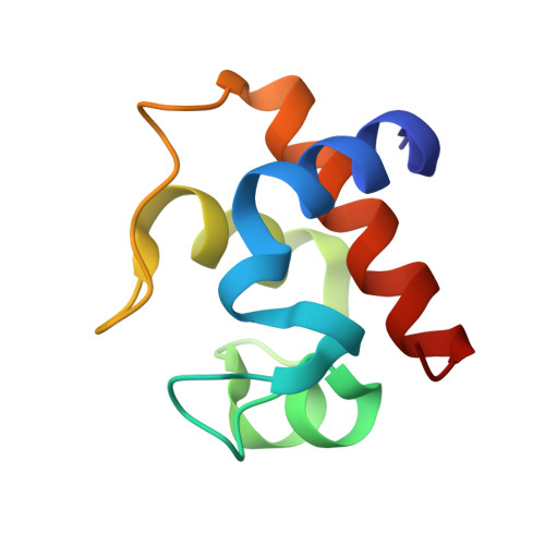
    3DMI

  • PubMed Abstract: 

    We determined for the first time the crystal structure of diatom cytochrome c(6) from Phaeodactylum tricornutum at 1.5 A resolution. The overall structure of the protein was classified as a class I c-type cytochrome. The physicochemical properties of the protein were examined by denaturation with guanidine hydrochloride and urea, and compared with those of other algal cytochrome c(6).


  • Organizational Affiliation

    Bio-Organic Chemistry Laboratory, Graduate School of Bioresource Sciences, Nihon University.


Macromolecules
Find similar proteins by:  (by identity cutoff)  |  3D Structure
Entity ID: 1
MoleculeChains Sequence LengthOrganismDetailsImage
cytochrome c688Phaeodactylum tricornutumMutation(s): 0 
Gene Names: pet J
UniProt
Find proteins for Q8GTB5 (Phaeodactylum tricornutum)
Explore Q8GTB5 
Go to UniProtKB:  Q8GTB5
Entity Groups  
Sequence Clusters30% Identity50% Identity70% Identity90% Identity95% Identity100% Identity
UniProt GroupQ8GTB5
Sequence Annotations
Expand
  • Reference Sequence
Small Molecules
Experimental Data & Validation

Experimental Data

  • Method: X-RAY DIFFRACTION
  • Resolution: 1.50 Å
  • R-Value Free: 0.202 
  • R-Value Work: 0.164 
  • R-Value Observed: 0.166 
  • Space Group: I 2 3
Unit Cell:
Length ( Å )Angle ( ˚ )
a = 80.385α = 90
b = 80.385β = 90
c = 80.385γ = 90
Software Package:
Software NamePurpose
REFMACrefinement
HKL-2000data collection
HKL-2000data reduction
SCALEPACKdata scaling
MOLREPphasing

Structure Validation

View Full Validation Report


Ligand Structure Quality Assessment 


Entry History 

Deposition Data

Revision History  (Full details and data files)

  • Version 1.0: 2009-03-17
    Type: Initial release
  • Version 1.1: 2011-07-13
    Changes: Version format compliance
  • Version 2.0: 2019-10-02
    Changes: Atomic model, Data collection, Derived calculations, Non-polymer description, Structure summary
  • Version 2.1: 2019-11-06
    Changes: Data collection, Database references
  • Version 2.2: 2023-11-01
    Changes: Data collection, Database references, Derived calculations, Refinement description