


Crystal structure of adenylate kinase variant AKlse1.
Bannen, R.M., Bae, E., McCoy, J.G., Phillips Jr., G.N.To be published.
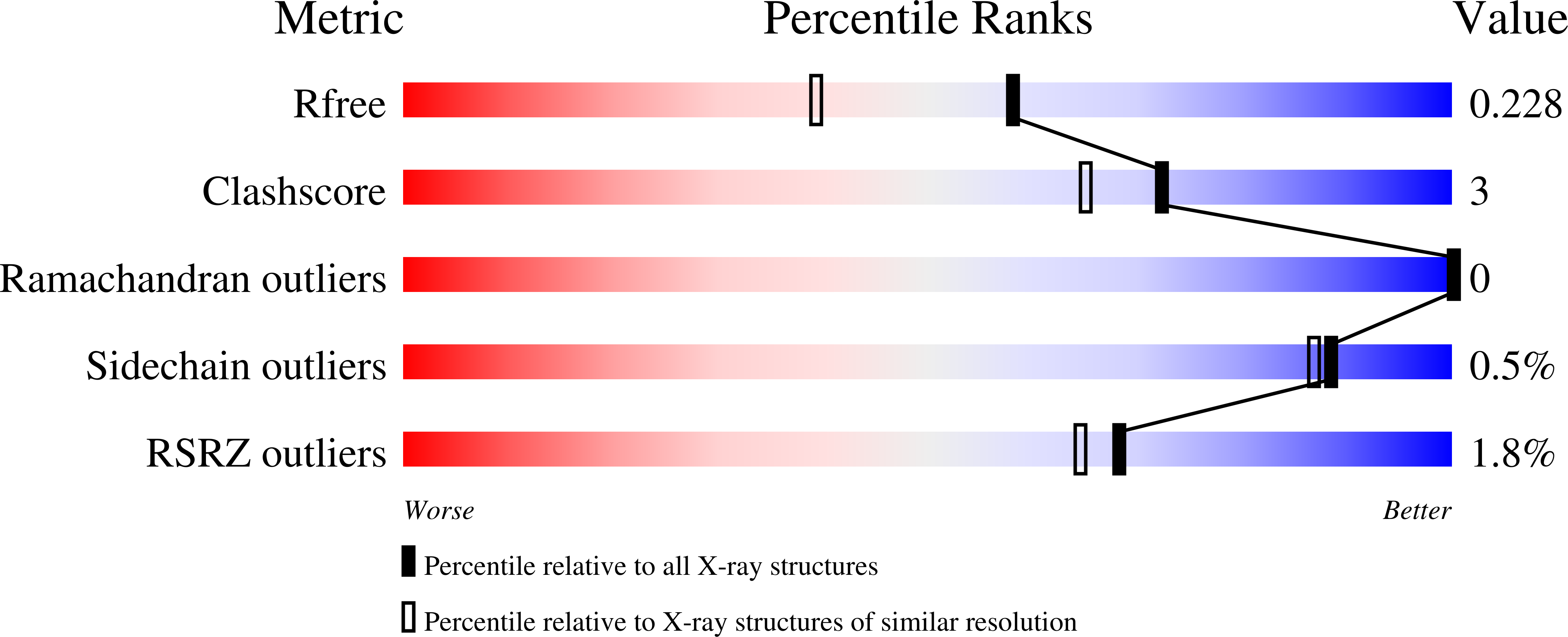
Experimental Data Snapshot
Starting Model: experimental
View more details
Entity ID: 1 | |||||
|---|---|---|---|---|---|
| Molecule | Chains | Sequence Length | Organism | Details | Image |
| Adenylate kinase | 217 | Bacillus subtilis | Mutation(s): 0 Gene Names: ADK EC: 2.7.4.3 | 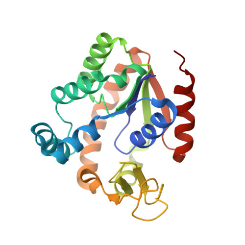 | |
UniProt | |||||
Find proteins for P16304 (Bacillus subtilis (strain 168)) Explore P16304 Go to UniProtKB: P16304 | |||||
Entity Groups | |||||
| Sequence Clusters | 30% Identity50% Identity70% Identity90% Identity95% Identity100% Identity | ||||
| UniProt Group | P16304 | ||||
Sequence AnnotationsExpand | |||||
| |||||
| Ligands 4 Unique | |||||
|---|---|---|---|---|---|
| ID | Chains | Name / Formula / InChI Key | 2D Diagram | 3D Interactions | |
| AP5 Query on AP5 | D [auth A] | BIS(ADENOSINE)-5'-PENTAPHOSPHATE C20 H29 N10 O22 P5 OIMACDRJUANHTJ-XPWFQUROSA-N |  | ||
| ZN Query on ZN | B [auth A] | ZINC ION Zn PTFCDOFLOPIGGS-UHFFFAOYSA-N |  | ||
| EDO Query on EDO | E [auth A], F [auth A], G [auth A] | 1,2-ETHANEDIOL C2 H6 O2 LYCAIKOWRPUZTN-UHFFFAOYSA-N |  | ||
| MG Query on MG | C [auth A] | MAGNESIUM ION Mg JLVVSXFLKOJNIY-UHFFFAOYSA-N |  | ||
| Length ( Å ) | Angle ( ˚ ) |
|---|---|
| a = 44.624 | α = 90 |
| b = 62.056 | β = 90 |
| c = 86.995 | γ = 90 |
| Software Name | Purpose |
|---|---|
| MOLREP | phasing |
| REFMAC | refinement |
| PDB_EXTRACT | data extraction |
| MAR345 | data collection |
| HKL-2000 | data reduction |
| HKL-2000 | data scaling |
RCSB PDB (citation) is hosted by
RCSB PDB is a member of the

