Mutating the tight-dimer interface of dihydrodipicolinate synthase disrupts the enzyme quaternary structure: toward a monomeric enzyme
Pearce, F.G., Dobson, R.C.J., Weber, A., Lane, L.A., McCammon, M.G., Squire, M.A., Perugini, M.A., Jameson, G.B., Robinson, C.V., Gerrard, J.A.(2008) Biochemistry 47: 12108-12117
- PubMed: 18937497
- DOI: https://doi.org/10.1021/bi801094t
- Primary Citation of Related Structures:
3DEN - PubMed Abstract:
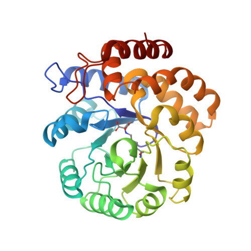
Dihydrodipicolinate synthase (DHDPS) is a tetrameric enzyme that is the first enzyme unique to the ( S)-lysine biosynthetic pathway in plants and bacteria. Previous studies have looked at the important role of Tyr107, an amino acid residue located at the tight-dimer interface between two monomers, in participating in a catalytic triad of residues during catalysis. In this study, we examine the importance of this residue in determining the quaternary structure of the DHDPS enzyme. The Tyr107 residue was mutated to tryptophan, and structural, biophysical, and kinetic studies were carried out on the mutant enzyme. These revealed that while the solid-state structure of the mutant enzyme was largely unchanged, as judged by X-ray crystallography, it exists as a mixture of primarily monomer and tetramer in solution, as determined by analytical ultracentrifugation, size-exclusion chromatography, and mass spectrometry. The catalytic ability of the DHDPS enzyme was reduced by the mutation, which also allowed the adventitious binding of alpha-ketoglutarate to the active site. A reduction in the apparent melting temperature of the mutant enzyme was observed. Thus, the tetrameric quaternary structure of DHDPS is critical to controlling specificity, heat stability, and intrinsic activity.
Organizational Affiliation:
School of Biological Sciences, University of Canterbury, Private Bag 4800, Christchurch 8020, New Zealand.