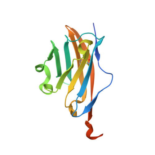
Demyelinating myelin oligodendrocyte glycoprotein-specific autoantibody response is focused on one dominant conformational epitope region in rodents
Breithaupt, C., Schafer, B., Pellkofer, H., Huber, R., Linington, C., Jacob, U.(2008) J Immunol 181: 1255-1263
- PubMed: 18606679
- DOI: https://doi.org/10.4049/jimmunol.181.2.1255
- Primary Citation of Related Structures:
3CSP - PubMed Abstract:
Conformational epitopes of myelin oligodendrocyte glycoprotein (MOG) provide a major target for demyelinating autoantibodies in experimental autoimmune encephalomyelitis and recent studies indicate that a similar situation may exist in multiple sclerosis. We recently solved the crystal structure of the extracellular domain of MOG (MOG(ex)) in complex with a Fab derived from the demyelinating mAb 8-18C5 and identified the conformational 8-18C5 epitope on MOG that is dominated by the surface exposed FG loop of MOG. To determine the importance of this epitope with regard to the polyclonal Ab response to MOG(ex) we investigated the effects of mutating His(103) and Ser(104), the two central amino acids of the FG loop, on Ab binding. Mutation of these two residues reduced binding of a panel of eight demyelinating conformation-dependent mAbs to <20% compared with binding to wild-type MOG(ex), whereas substitution of amino acids that do not contribute to the 8-18C5 epitope had only a minor effect on Ab binding. The same restriction was observed for the polyclonal MOG-specific Ab response of MOG DNA-vaccinated BALB/c and SJL/J mice. Our data demonstrate that the pathogenic anti-MOG Ab response primarily targets one immunodominant region centered at the FG loop of MOG. Comparison of the structure of MOG(ex) with the structures of related IgV-like domains yields a possible explanation for the focused Ab response.
Organizational Affiliation:
Max-Planck-Institut für Biochemie, Abteilung Strukturforschung, Emeritusgruppe Huber, Martinsried, Germany. constanze.breithaupt@biochemtech.uni-halle.de