Contributions of all 20 amino acids at site 96 to the stability and structure of T4 lysozyme.
Mooers, B.H., Baase, W.A., Wray, J.W., Matthews, B.W.(2009) Protein Sci 18: 871-880
- PubMed: 19384988
- DOI: https://doi.org/10.1002/pro.94
- Primary Citation of Related Structures:
3C7W, 3C7Y, 3C7Z, 3C80, 3C81, 3C82, 3C83, 3C8Q, 3C8R, 3C8S, 3CDO, 3CDQ, 3CDR, 3CDT, 3CDV, 3FI5 - PubMed Abstract:
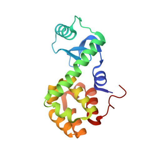
To try to resolve the loss of stability in the temperature-sensitive mutant of T4 lysozyme, Arg 96 --> His, all of the remaining 18 naturally occurring amino acids were substituted at site 96. Also, in response to suggestions that the charged residues Lys85 and Asp89, which are 5-8 A away, may have important effects, each of these amino acids was replaced with alanine. Crystal structures were determined for many of the variants. With the exception of the tryptophan and valine mutants R96W and R96V, the crystallographic analysis shows that the substituted side chain following the path of Arg96 in wildtype (WT). The melting temperatures of the variants decrease by up to approximately 16 degrees C with WT being most stable. There are two site 96 replacements, with lysine or glutamine, that leave the stability close to that of WT. The only element that the side chains of these residues have in common with the WT arginine is the set of three carbon atoms at the C(alpha), C(beta), and C(gamma) positions. Although each side chain is long and flexible with a polar group at the distal position, the details of the hydrogen bonding to the rest of the protein differ in each case. Also, the glutamine replacement lacks a positive charge. This shows that there is some adaptability in achieving full stabilization at this site. At the other extreme, to be maximally destabilizing a mutation at site 96 must not only eliminate favorable interactions but also introduce an unfavorable element such as steric strain or a hydrogen-bonding group that remains unsatisfied. Overall, the study highlights the essential need for atomic resolution site-specific structural information to understand and to predict the stability of mutant proteins. It can be very misleading to simply assume that conservative amino acid substitutions cause small changes in stability, whereas large stability changes are associated with nonconservative replacements.
Organizational Affiliation:
Institute of Molecular Biology, Howard Hughes Medical Institute, University of Oregon, Eugene, Oregon 97403-1229, USA.