3C7N

Structure of the Hsp110:Hsc70 Nucleotide Exchange Complex


Experimental Data Snapshot

  • Method: X-RAY DIFFRACTION
  • Resolution: 3.12 Å
  • R-Value Free: 0.283 
  • R-Value Work: 0.224 
  • R-Value Observed: 0.227 

wwPDB Validation   3D Report Full Report


Ligand Structure Quality Assessment 


This is version 1.3 of the entry. See complete history


Literature

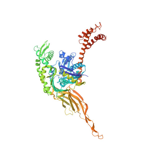
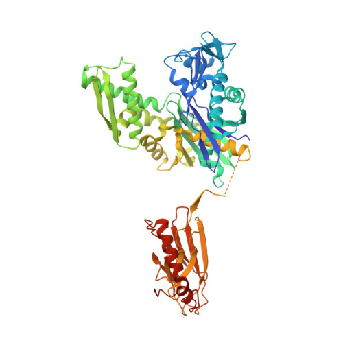
Structure of the Hsp110:Hsc70 nucleotide exchange machine

Schuermann, J.P.Jiang, J.Cuellar, J.Llorca, O.Wang, L.Gimenez, L.E.Jin, S.Taylor, A.B.Demeler, B.Morano, K.A.Hart, P.J.Valpuesta, J.M.Lafer, E.M.Sousa, R.

(2008) Mol Cell 31: 232-243

  • DOI: https://doi.org/10.1016/j.molcel.2008.05.006
  • Primary Citation of Related Structures:  
    3C7N

  • PubMed Abstract: 

    Hsp70s mediate protein folding, translocation, and macromolecular complex remodeling reactions. Their activities are regulated by proteins that exchange ADP for ATP from the nucleotide-binding domain (NBD) of the Hsp70. These nucleotide exchange factors (NEFs) include the Hsp110s, which are themselves members of the Hsp70 family. We report the structure of an Hsp110:Hsc70 nucleotide exchange complex. The complex is characterized by extensive protein:protein interactions and symmetric bridging interactions between the nucleotides bound in each partner protein's NBD. An electropositive pore allows nucleotides to enter and exit the complex. The role of nucleotides in complex formation and dissociation, and the effects of the protein:protein interactions on nucleotide exchange, can be understood in terms of the coupled effects of the nucleotides and protein:protein interactions on the open-closed isomerization of the NBDs. The symmetrical interactions in the complex may model other Hsp70 family heterodimers in which two Hsp70s reciprocally act as NEFs.


  • Organizational Affiliation

    Department of Biochemistry, University of Texas Health Science Center, San Antonio, TX 78229-3900, USA.


Macromolecules
Find similar proteins by:  (by identity cutoff)  |  3D Structure
Entity ID: 1
MoleculeChains Sequence LengthOrganismDetailsImage
Heat shock protein homolog SSE1668Saccharomyces cerevisiaeMutation(s): 0 
Gene Names: SSE1MSI3
UniProt
Find proteins for P32589 (Saccharomyces cerevisiae (strain ATCC 204508 / S288c))
Explore P32589 
Go to UniProtKB:  P32589
Entity Groups  
Sequence Clusters30% Identity50% Identity70% Identity90% Identity95% Identity100% Identity
UniProt GroupP32589
Sequence Annotations
Expand
  • Reference Sequence
Find similar proteins by:  (by identity cutoff)  |  3D Structure
Entity ID: 2
MoleculeChains Sequence LengthOrganismDetailsImage
Heat shock cognate554Bos taurusMutation(s): 0 
Gene Names: HSPA8HSC70
UniProt
Find proteins for P19120 (Bos taurus)
Explore P19120 
Go to UniProtKB:  P19120
Entity Groups  
Sequence Clusters30% Identity50% Identity70% Identity90% Identity95% Identity100% Identity
UniProt GroupP19120
Sequence Annotations
Expand
  • Reference Sequence
Small Molecules
Ligands 5 Unique
IDChains Name / Formula / InChI Key2D Diagram3D Interactions
ADP
Query on ADP

Download Ideal Coordinates CCD File 
H [auth A],
N [auth B]
ADENOSINE-5'-DIPHOSPHATE
C10 H15 N5 O10 P2
XTWYTFMLZFPYCI-KQYNXXCUSA-N
SO4
Query on SO4

Download Ideal Coordinates CCD File 
D [auth A],
E [auth A],
F [auth A],
M [auth B]
SULFATE ION
O4 S
QAOWNCQODCNURD-UHFFFAOYSA-L
BEF
Query on BEF

Download Ideal Coordinates CCD File 
G [auth A]BERYLLIUM TRIFLUORIDE ION
Be F3
OGIAHMCCNXDTIE-UHFFFAOYSA-K
CL
Query on CL

Download Ideal Coordinates CCD File 
I [auth B],
J [auth B],
K [auth B]
CHLORIDE ION
Cl
VEXZGXHMUGYJMC-UHFFFAOYSA-M
MG
Query on MG

Download Ideal Coordinates CCD File 
C [auth A],
L [auth B]
MAGNESIUM ION
Mg
JLVVSXFLKOJNIY-UHFFFAOYSA-N
Binding Affinity Annotations 
IDSourceBinding Affinity
ADP BindingDB:  3C7N Kd: min: 320, max: 324 (nM) from 2 assay(s)
IC50: 280 (nM) from 1 assay(s)
PDBBind:  3C7N Kd: 200 (nM) from 1 assay(s)
Experimental Data & Validation

Experimental Data

  • Method: X-RAY DIFFRACTION
  • Resolution: 3.12 Å
  • R-Value Free: 0.283 
  • R-Value Work: 0.224 
  • R-Value Observed: 0.227 
  • Space Group: P 21 21 2
Unit Cell:
Length ( Å )Angle ( ˚ )
a = 123.533α = 90
b = 169.501β = 90
c = 87.724γ = 90
Software Package:
Software NamePurpose
DENZOdata reduction
SCALEPACKdata scaling
MOLREPphasing
PHENIXrefinement
PDB_EXTRACTdata extraction
HKL-2000data collection
HKL-2000data reduction
HKL-2000data scaling

Structure Validation

View Full Validation Report


Ligand Structure Quality Assessment 


Entry History 

Deposition Data

Revision History  (Full details and data files)

  • Version 1.0: 2008-05-27
    Type: Initial release
  • Version 1.1: 2011-07-13
    Changes: Version format compliance
  • Version 1.2: 2017-10-25
    Changes: Refinement description, Source and taxonomy
  • Version 1.3: 2024-02-21
    Changes: Data collection, Database references, Derived calculations