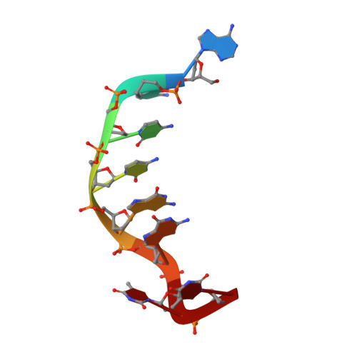
3C2J

Crystal structure analysis of trioxacarcin A covalently bound to d(AACCGGTT)

  • Classification: DNA
  • Mutation(s): No 

  • Deposited: 2008-01-25 Released: 2008-06-03 
  • Deposition Author(s): Pfoh, R., Sheldrick, G.M.

Experimental Data Snapshot

  • Method: X-RAY DIFFRACTION
  • Resolution: 1.78 Å
  • R-Value Free: 0.265 
  • R-Value Work: 0.220 
  • R-Value Observed: 0.222 

wwPDB Validation   3D Report Full Report


Ligand Structure Quality Assessment 


This is version 1.2 of the entry. See complete history


Literature

Crystal structure of trioxacarcin A covalently bound to DNA

Pfoh, R.Laatsch, H.Sheldrick, G.M.

(2008) Nucleic Acids Res 36: 3508-3514

  • DOI: https://doi.org/10.1093/nar/gkn245
  • Primary Citation of Related Structures:  
    3C2J

  • PubMed Abstract: 

    We report a crystal structure that shows an antibiotic that extracts a nucleobase from a DNA molecule 'caught in the act' after forming a covalent bond but before departing with the base. The structure of trioxacarcin A covalently bound to double-stranded d(AACCGGTT) was determined to 1.78 A resolution by MAD phasing employing brominated oligonucleotides. The DNA-drug complex has a unique structure that combines alkylation (at the N7 position of a guanine), intercalation (on the 3'-side of the alkylated guanine), and base flip-out. An antibiotic-induced flipping-out of a single, nonterminal nucleobase from a DNA duplex was observed for the first time in a crystal structure.


  • Organizational Affiliation

    Lehrstuhl für Strukturchemie, Georg-August-Universität, Tammannstr. 4, 37077 Göttingen, Germany.


Macromolecules

Find similar nucleic acids by:  Sequence   |   3D Structure  

Entity ID: 1
MoleculeChains LengthOrganismImage
DNA (5'-D(*DAP*DAP*DCP*DCP*DGP*DGP*DTP*DT)-3')
A, B
8N/A
Sequence Annotations
Expand
  • Reference Sequence
Experimental Data & Validation

Experimental Data

  • Method: X-RAY DIFFRACTION
  • Resolution: 1.78 Å
  • R-Value Free: 0.265 
  • R-Value Work: 0.220 
  • R-Value Observed: 0.222 
  • Space Group: P 41 2 2
Unit Cell:
Length ( Å )Angle ( ˚ )
a = 37.6α = 90
b = 37.6β = 90
c = 91.62γ = 90
Software Package:
Software NamePurpose
REFMACrefinement
PDB_EXTRACTdata extraction
MAR345dtbdata collection
HKL-2000data reduction
SADABSdata scaling
SHELXDEphasing

Structure Validation

View Full Validation Report


Ligand Structure Quality Assessment 


Entry History 

Deposition Data

Revision History  (Full details and data files)

  • Version 1.0: 2008-06-03
    Type: Initial release
  • Version 1.1: 2011-07-13
    Changes: Advisory, Refinement description, Version format compliance
  • Version 1.2: 2024-03-13
    Changes: Data collection, Database references, Derived calculations