Crystal structure of Dioclea violacea lectin and a comparative study of vasorelaxant properties with Dioclea rostrata lectin
Bezerra, M.J.B., Rodrigues, N.V.F.C., Pires, A.F., Bezerra, G.A., Nobre, C.B., Alencar, K.L.L., Soares, P.M.G., Nascimento, K.S., Nagano, C.S., Martins, J.L., Gruber, K., Sampaio, A.H., Delatorre, P., Rocha, B.A.M., Assreuy, A.M.S., Cavada, B.S.(2013) Int J Biochem Cell Biol 45: 807-815
- PubMed: 23353644
- DOI: https://doi.org/10.1016/j.biocel.2013.01.012
- Primary Citation of Related Structures:
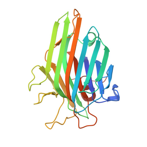
3AX4 - PubMed Abstract:
Lectins from Diocleinae subtribe belong to the family of legume lectins and are characterized by high identity between their amino acids sequences. It has been shown that punctual differences in amino acid sequences, such as one single amino acid or an alternative conformation, represent changes in biological activities caused by these lectins. Therefore, a more detailed understanding of three-dimensional structures of these proteins is essential for accurate analyzing the relationship between structure and function. In this study lectins purified from the seeds of Dioclea violacea (DVL) and Dioclea rostrata (DRL) were compared with regard to crystal structure and vasorelaxant properties. Differences in structure of lectins were found to be reflected in differences in vasorelaxant effects based on their high specificity and selectivity for cell glycans. Binding activity was related to the position of specific residues in the carbohydrate recognition domain (CRD). DVL complexed structure was solved by X-ray crystallography and was compared to native DVL and DRL. Therefore, DVL was co-crystallized with X-Man, and a molecular modeling with X-Man complexed with DVL was done to compare the complexed and native forms adjusted fit. The relatively narrow and deep CRD in DVL promotes little interaction with carbohydrates; in contrast, the wider and shallower CRD in DRL favors interaction. This seems to explain differences in the level of relaxation induced by DVL (43%) and DRL (96%) in rat aortic rings.
Organizational Affiliation:
Departamento de Bioquímica e Biologia Molecular, Universidade Federal do Ceará, Av. Mister Hull s/n, Bloco 907, Box 6043, 60440-970, Fortaleza, Ceará, Brazil.