3AM1

Crystal structure of O-Phosphoseryl-tRNA kinase complexed with anticodon-stem/loop truncated tRNA(Sec)


Experimental Data Snapshot

  • Method: X-RAY DIFFRACTION
  • Resolution: 2.40 Å
  • R-Value Free: 0.292 
  • R-Value Work: 0.229 
  • R-Value Observed: 0.232 

wwPDB Validation   3D Report Full Report


Ligand Structure Quality Assessment 


This is version 1.3 of the entry. See complete history


Literature

C-terminal domain of archaeal O-phosphoseryl-tRNA kinase displays large-scale motion to bind the 7-bp D-stem of archaeal tRNA(Sec)

Sherrer, R.L.Araiso, Y.Aldag, C.Ishitani, R.Ho, J.M.L.Soll, D.Nureki, O.

(2011) Nucleic Acids Res 39: 1034-1041

  • DOI: https://doi.org/10.1093/nar/gkq845
  • Primary Citation of Related Structures:  
    3AM1

  • PubMed Abstract: 

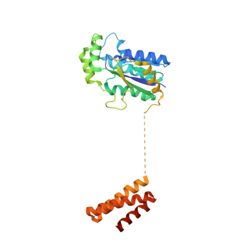
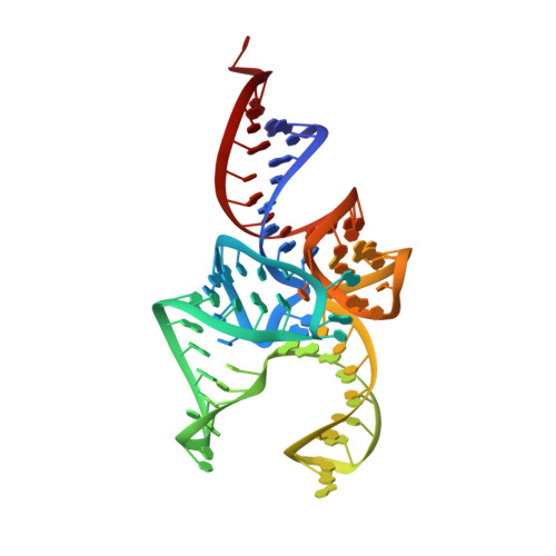
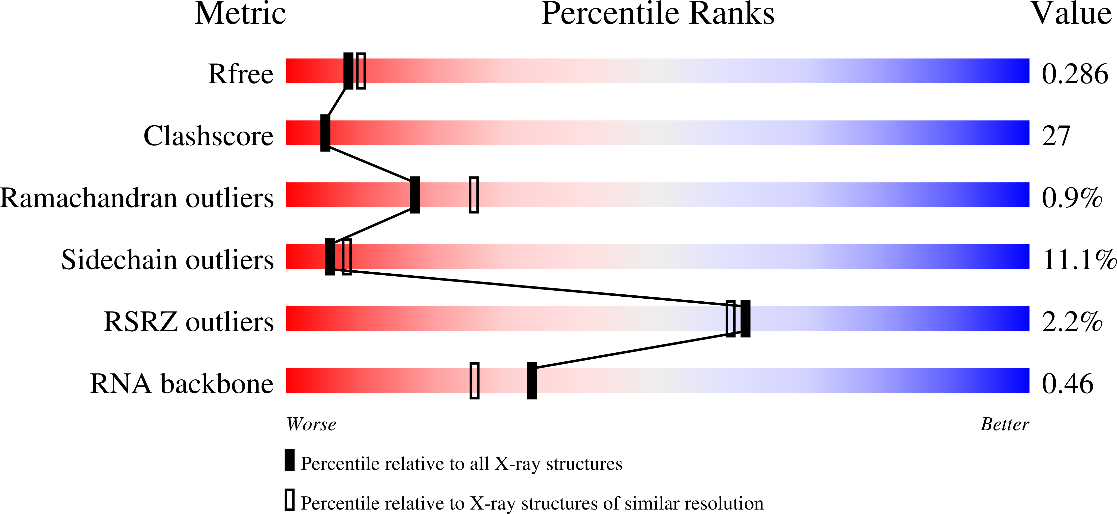
    O-Phosphoseryl-tRNA kinase (PSTK) is the key enzyme in recruiting selenocysteine (Sec) to the genetic code of archaea and eukaryotes. The enzyme phosphorylates Ser-tRNA(Sec) to produce O-phosphoseryl-tRNA(Sec) (Sep-tRNA(Sec)) that is then converted to Sec-tRNA(Sec) by Sep-tRNA:Sec-tRNA synthase. Earlier we reported the structure of the Methanocaldococcus jannaschii PSTK (MjPSTK) complexed with AMPPNP. This study presents the crystal structure (at 2.4-Å resolution) of MjPSTK complexed with an anticodon-stem/loop truncated tRNA(Sec) (Mj*tRNA(Sec)), a good enzyme substrate. Mj*tRNA(Sec) is bound between the enzyme's C-terminal domain (CTD) and N-terminal kinase domain (NTD) that are connected by a flexible 11 amino acid linker. Upon Mj*tRNA(Sec) recognition the CTD undergoes a 62-Å movement to allow proper binding of the 7-bp D-stem. This large reorganization of the PSTK quaternary structure likely provides a means by which the unique tRNA(Sec) species can be accurately recognized with high affinity by the translation machinery. However, while the NTD recognizes the tRNA acceptor helix, shortened versions of MjPSTK (representing only 60% of the original size, in which the entire CTD, linker loop and an adjacent NTD helix are missing) are still active in vivo and in vitro, albeit with reduced activity compared to the full-length enzyme.


  • Organizational Affiliation

    Department of Molecular Biophysics and Biochemistry, Yale University, New Haven, Connecticut 06520-8114, USA.


Macromolecules

Find similar proteins by:  (by identity cutoff)  |  3D Structure
Entity ID: 1
MoleculeChains Sequence LengthOrganismDetailsImage
L-seryl-tRNA(Sec) kinase260Methanocaldococcus jannaschiiMutation(s): 0 
Gene Names: pstKMJ1538
EC: 2.7.1.164
UniProt
Find proteins for Q58933 (Methanocaldococcus jannaschii (strain ATCC 43067 / DSM 2661 / JAL-1 / JCM 10045 / NBRC 100440))
Explore Q58933 
Go to UniProtKB:  Q58933
Entity Groups  
Sequence Clusters30% Identity50% Identity70% Identity90% Identity95% Identity100% Identity
UniProt GroupQ58933
Sequence Annotations
Expand
  • Reference Sequence
Find similar nucleic acids by:  (by identity cutoff)  |  3D Structure
Entity ID: 2
MoleculeChains LengthOrganismImage
ASL-truncated tRNA81N/A
Sequence Annotations
Expand
  • Reference Sequence
Small Molecules
Ligands 2 Unique
IDChains Name / Formula / InChI Key2D Diagram3D Interactions
ATP
Query on ATP

Download Ideal Coordinates CCD File 
C [auth A]ADENOSINE-5'-TRIPHOSPHATE
C10 H16 N5 O13 P3
ZKHQWZAMYRWXGA-KQYNXXCUSA-N
MG
Query on MG

Download Ideal Coordinates CCD File 
D [auth A]MAGNESIUM ION
Mg
JLVVSXFLKOJNIY-UHFFFAOYSA-N
Experimental Data & Validation

Experimental Data

  • Method: X-RAY DIFFRACTION
  • Resolution: 2.40 Å
  • R-Value Free: 0.292 
  • R-Value Work: 0.229 
  • R-Value Observed: 0.232 
  • Space Group: P 31 1 2
Unit Cell:
Length ( Å )Angle ( ˚ )
a = 46.749α = 90
b = 46.749β = 90
c = 459.712γ = 120
Software Package:
Software NamePurpose
SCALEPACKdata scaling
PHENIXrefinement
PDB_EXTRACTdata extraction
HKL-2000data reduction
HKL-2000data scaling
MOLREPphasing

Structure Validation

View Full Validation Report


Ligand Structure Quality Assessment 


Entry History 

Deposition Data

Revision History  (Full details and data files)

  • Version 1.0: 2010-12-29
    Type: Initial release
  • Version 1.1: 2011-07-13
    Changes: Version format compliance
  • Version 1.2: 2014-01-22
    Changes: Database references
  • Version 1.3: 2023-11-01
    Changes: Data collection, Database references, Derived calculations, Refinement description