3ALW

Crystal structure of the measles virus hemagglutinin bound to its cellular receptor SLAM (Form I, MV-H-SLAM(N102H/R108Y) fusion)


Experimental Data Snapshot

  • Method: X-RAY DIFFRACTION
  • Resolution: 3.55 Å
  • R-Value Free: 0.283 
  • R-Value Work: 0.250 
  • R-Value Observed: 0.250 

wwPDB Validation   3D Report Full Report


Ligand Structure Quality Assessment 


This is version 1.4 of the entry. See complete history


Literature

Structure of the measles virus hemagglutinin bound to its cellular receptor SLAM

Hashiguchi, T.Ose, T.Kubota, M.Maita, N.Kamishikiryo, J.Maenaka, K.Yanagi, Y.

(2011) Nat Struct Mol Biol 18: 135-141

  • DOI: https://doi.org/10.1038/nsmb.1969
  • Primary Citation of Related Structures:  
    3ALW, 3ALX, 3ALZ

  • PubMed Abstract: 

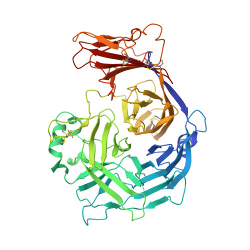
    Measles virus, a major cause of childhood morbidity and mortality worldwide, predominantly infects immune cells using signaling lymphocyte activation molecule (SLAM) as a cellular receptor. Here we present crystal structures of measles virus hemagglutinin (MV-H), the receptor-binding glycoprotein, in complex with SLAM. The MV-H head domain binds to a β-sheet of the membrane-distal ectodomain of SLAM using the side of its β-propeller fold. This is distinct from attachment proteins of other paramyxoviruses that bind receptors using the top of their β-propeller. The structure provides templates for antiviral drug design, an explanation for the effectiveness of the measles virus vaccine, and a model of the homophilic SLAM-SLAM interaction involved in immune modulations. Notably, the crystal structures obtained show two forms of the MV-H-SLAM tetrameric assembly (dimer of dimers), which may have implications for the mechanism of fusion triggering.


  • Organizational Affiliation

    Department of Virology, Faculty of Medicine, Kyushu University, Fukuoka, Japan.


Macromolecules
Find similar proteins by:  (by identity cutoff)  |  3D Structure
Entity ID: 1
MoleculeChains Sequence LengthOrganismDetailsImage
Hemagglutinin, CDw150559Measles morbillivirussynthetic constructSaguinus oedipus
This entity is chimeric
Mutation(s): 2 
Gene Names: HemagglutininCD150
UniProt
Find proteins for P08362 (Measles virus (strain Edmonston))
Explore P08362 
Go to UniProtKB:  P08362
Find proteins for Q9GJT3 (Saguinus oedipus)
Explore Q9GJT3 
Go to UniProtKB:  Q9GJT3
Entity Groups  
Sequence Clusters30% Identity50% Identity70% Identity90% Identity95% Identity100% Identity
UniProt GroupsP08362Q9GJT3
Sequence Annotations
Expand
  • Reference Sequence
Small Molecules
Ligands 1 Unique
IDChains Name / Formula / InChI Key2D Diagram3D Interactions
NAG
Query on NAG

Download Ideal Coordinates CCD File 
B [auth A],
C [auth A]
2-acetamido-2-deoxy-beta-D-glucopyranose
C8 H15 N O6
OVRNDRQMDRJTHS-FMDGEEDCSA-N
Experimental Data & Validation

Experimental Data

  • Method: X-RAY DIFFRACTION
  • Resolution: 3.55 Å
  • R-Value Free: 0.283 
  • R-Value Work: 0.250 
  • R-Value Observed: 0.250 
  • Space Group: P 63 2 2
Unit Cell:
Length ( Å )Angle ( ˚ )
a = 209.912α = 90
b = 209.912β = 90
c = 180.538γ = 120
Software Package:
Software NamePurpose
ADSCdata collection
PHASERphasing
CNSrefinement
HKL-2000data reduction
HKL-2000data scaling

Structure Validation

View Full Validation Report


Ligand Structure Quality Assessment 


Entry History 

Deposition Data

Revision History  (Full details and data files)

  • Version 1.0: 2011-01-12
    Type: Initial release
  • Version 1.1: 2011-07-13
    Changes: Version format compliance
  • Version 1.2: 2017-06-28
    Changes: Source and taxonomy
  • Version 1.3: 2020-07-29
    Type: Remediation
    Reason: Carbohydrate remediation
    Changes: Data collection, Database references, Derived calculations, Structure summary
  • Version 1.4: 2023-11-01
    Changes: Data collection, Database references, Refinement description, Structure summary