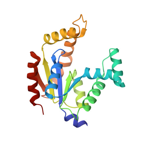
Refined structure of porcine cytosolic adenylate kinase at 2.1 A resolution.
Dreusicke, D., Karplus, P.A., Schulz, G.E.(1988) J Mol Biol 199: 359-371
- PubMed: 2832612
- DOI: https://doi.org/10.1016/0022-2836(88)90319-1
- Primary Citation of Related Structures:
3ADK - PubMed Abstract:
The crystal structure of porcine cytosolic adenylate kinase has been established at 2.1 A resolution using a restrained least-squares refinement method. Based on 11,251 independent reflections of better than 10 A resolution, a final R-factor of 19.3% was obtained with a model obeying standard geometry within 0.026 A in bond lengths and 3.3 degrees in bond angles. In comparison with the previous structure at 3 A resolution, there is a significant improvement. The high resolution structure has been used to rationalize the strictly conserved residues in the adenylate kinase family. Among these is the glycine-rich loop, which forms a giant anion hole accommodating a sulfate ion which mimics a phosphoryl group of a substrate. Such a structure seems to occur in a large group of mononucleotide binding proteins. Moreover, a conserved cis-proline has been detected in the active center. A structural comparison with the complex between adenylate kinase from yeast and a substrate-analog at medium resolution indicates that this kinase performs appreciable mechanical movements during a catalytic cycle. The reported structure presumably represents an open form of the enzyme, similar to that in solution in the absence of substrates. However, since there are large intermolecular contacts in the crystal, some deviation from the solution structure has to be expected.
Organizational Affiliation:
Institut für Organische Chemie und Biochemie der Universität, Freiburg i.Br., Federal Republic of Germany.


