360D

STRUCTURE OF 2,5-BIS{[4-(N-ETHYLAMIDINO)PHENYL]}FURAN COMPLEXED TO 5'-D(CPGPCPGPAPAPTPTPCPGPCPG)-3'. A MINOR GROOVE DRUG COMPLEX, SHOWING PATTERNS OF GROOVE HYDRATION


Experimental Data Snapshot

  • Method: X-RAY DIFFRACTION
  • Resolution: 1.85 Å
  • R-Value Work: 0.166 

wwPDB Validation   3D Report Full Report


Ligand Structure Quality Assessment 


This is version 1.6 of the entry. See complete history


Literature

Visualisation of extensive water ribbons and networks in a DNA minor-groove drug complex.

Guerri, A.Simpson, I.J.Neidle, S.

(1998) Nucleic Acids Res 26: 2873-2878

  • DOI: https://doi.org/10.1093/nar/26.12.2873
  • Primary Citation of Related Structures:  
    360D

  • PubMed Abstract: 

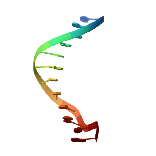
    The crystal structure is reported of a complex between an ethyl derivative of the minor-groove drug furamidine and the dodecanucleotide duplex d(CGCGAATTCGCG)2, which has been refined to 1.85 A resolution and an R factor of 16.6% for data collected at -173 degreesC. An exceptionally large number (220) of water molecules have been located. The majority of these occur in the first coordination shell of solvation. There are extensive networks of connected waters, both in the major and minor grooves. In particular, there are 21 water molecules associated with the minor-groove drug, via hydrogen bonds from the four charged nitrogen atoms. One cluster of four waters is situated in the groove itself; the majority are on the outer edge of the groove, and serve to bridge between the outward-directed drug nitrogen atoms and backbone phosphate oxygen atoms. These bridges are both intra- and inter-strand, with the net effect that the outer edge of the drug molecule is covered by ribbons of water molecules.


  • Organizational Affiliation

    The CRC Biomolecular Structure Unit, The Institute of Cancer Research, Sutton, Surrey SM2 5NG, UK.


Macromolecules

Find similar nucleic acids by:  Sequence   |   3D Structure  

Entity ID: 1
MoleculeChains LengthOrganismImage
DNA (5'-D(*CP*GP*CP*GP*AP*AP*TP*TP*CP*GP*CP*G)-3')
A, B
12synthetic construct
Sequence Annotations
Expand
  • Reference Sequence
Small Molecules
Ligands 2 Unique
IDChains Name / Formula / InChI Key2D Diagram3D Interactions
BPF
Query on BPF

Download Ideal Coordinates CCD File 
D [auth B]N,N'-(furan-2,5-diylbis{benzene-4,1-diyl[(Z)-aminomethylylidene]})diethanaminium
C22 H26 N4 O
UDRBFQQQLONWPG-UHFFFAOYSA-P
MG
Query on MG

Download Ideal Coordinates CCD File 
C [auth A]MAGNESIUM ION
Mg
JLVVSXFLKOJNIY-UHFFFAOYSA-N
Experimental Data & Validation

Experimental Data

  • Method: X-RAY DIFFRACTION
  • Resolution: 1.85 Å
  • R-Value Work: 0.166 
  • Space Group: P 21 21 21
Unit Cell:
Length ( Å )Angle ( ˚ )
a = 24.24α = 90
b = 39.94β = 90
c = 65.88γ = 90
Software Package:
Software NamePurpose
SHELXL-97refinement
DENZOdata reduction
SCALEPACKdata scaling

Structure Validation

View Full Validation Report


Ligand Structure Quality Assessment 


Entry History 

Deposition Data

Revision History  (Full details and data files)

  • Version 1.0: 1998-07-16
    Type: Initial release
  • Version 1.1: 2008-05-22
    Changes: Version format compliance
  • Version 1.2: 2011-07-13
    Changes: Version format compliance
  • Version 1.3: 2012-04-25
    Changes: Non-polymer description
  • Version 1.4: 2018-03-07
    Changes: Data collection
  • Version 1.5: 2020-04-08
    Changes: Structure summary
  • Version 1.6: 2023-08-02
    Changes: Database references, Derived calculations, Refinement description