Determination by MAD-DM of the structure of the DNA duplex d[ACGTACG(5-BrU)]2 at 1.46 A and 100 K.
Todd, A.K., Adams, A., Powell, H.R., Wilcock, D.J., Thorpe, J.H., Lausi, A., Zanini, F., Wakelin, L.P., Cardin, C.J.(1999) Acta Crystallogr D Biol Crystallogr 55: 729-735
- PubMed: 10089302
- DOI: https://doi.org/10.1107/s090744499801261x
- Primary Citation of Related Structures:
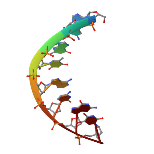
344D - PubMed Abstract:
A four-wavelength MAD experiment on a new brominated octanucleotide is reported here. d[ACGTACG(5-BrU)], C77H81BrN30O32P7, Mr (DNA) = 2235, tetragonal, P43212 (No. 96), a = 43.597, c = 26.268 A, V = 49927.5 A3, Z = 8, T = 100 K, R = 10.91% for 4312 reflections between 15.0 and 1.46 A resolution. The self-complementary brominated octanucleotide d[ACGTACG(5-BrU)]2 has been crystallized and data measured to 1.45 A at both 293 K and a second crystal flash frozen at 100 K. The latter data collection was carried out to the same resolution at the four wavelengths 0.9344, 0.9216, 0.9208 and 0.9003 A, around the Br K edge at 0.92 A and the structure determined from a map derived from a MAD data analysis using pseudo-MIR methodology, as implemented in the program MLPHARE. This is one of the first successful MAD phasing experiments carried out at Sincrotrone Elettra in Trieste, Italy. The structure was refined using the data measured at 0.9003 A, anisotropic temperature factors and the restrained least-squares refinement implemented in the program SHELX96, and the helical parameters are compared with those previously determined for the isomorphous d(ACGTACGT)2 analogue. The asymmetric unit consists of a single strand of octamer with 96 water molecules. No countercations were located. The A-DNA helix geometry obtained has been analysed using the CURVES program.
Organizational Affiliation:
Department of Chemistry, The University of Reading, Whiteknights, Reading RG6 6AD, England.