2ZQN

Crystal structure of the earthworm R-type lectin C-half in complex with Lactose


Experimental Data Snapshot

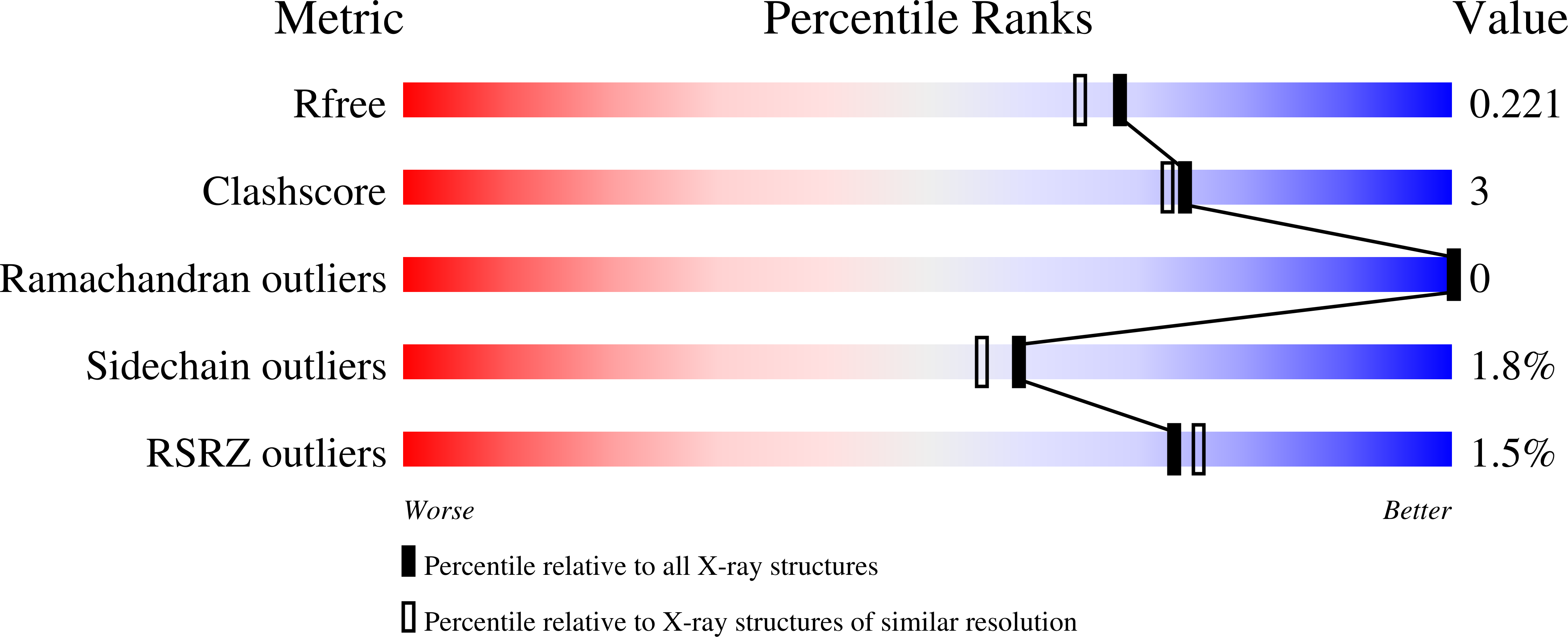
  • Method: X-RAY DIFFRACTION
  • Resolution: 1.90 Å
  • R-Value Free: 0.225 
  • R-Value Work: 0.176 
  • R-Value Observed: 0.179 

wwPDB Validation   3D Report Full Report


This is version 2.1 of the entry. See complete history


Literature

Sugar-complex structures of the C-half domain of the galactose-binding lectin EW29 from the earthworm Lumbricus terrestris

Suzuki, R.Kuno, A.Hasegawa, T.Hirabayashi, J.Kasai, K.Momma, M.Fujimoto, Z.

(2009) Acta Crystallogr D Biol Crystallogr 65: 49-57

  • DOI: https://doi.org/10.1107/S0907444908037451
  • Primary Citation of Related Structures:  
    2ZQN, 2ZQO

  • PubMed Abstract: 

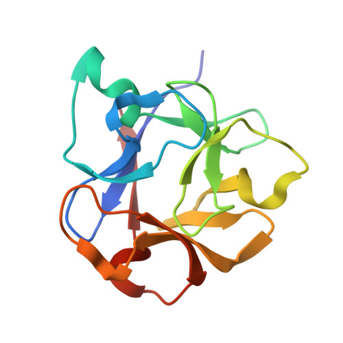
    R-type lectins are one of the most prominent types of lectin; they exist ubiquitously in nature and mainly bind to the galactose unit of sugar chains. The galactose-binding lectin EW29 from the earthworm Lumbricus terrestris belongs to the R-type lectin family as represented by the plant lectin ricin. It shows haemagglutination activity and is composed of a single peptide chain that includes two homologous domains: N-terminal and C-terminal domains. A truncated mutant of EW29 comprising the C-terminal domain (rC-half) has haemagglutination activity by itself. In order to clarify how rC-half recognizes ligands and shows haemagglutination activity, X-ray crystal structures of rC-half in complex with D-lactose and N-acetyl-D-galactosamine have been determined. The structure of rC-half is similar to that of the ricin B chain and consists of a beta-trefoil fold; the fold is further divided into three similar subdomains referred to as subdomains alpha, beta and gamma, which are gathered around the pseudo-threefold axis. The structures of sugar complexes demonstrated that subdomains alpha and gamma of rC-half bind terminal galactosyl and N-acetylgalactosaminyl glycans. The sugar-binding properties are common to both ligands in both subdomains and are quite similar to those of ricin B chain-lactose complexes. These results indicate that the C-terminal domain of EW29 uses these two galactose-binding sites for its function as a single-domain-type haemagglutinin.


  • Organizational Affiliation

    Protein Research Unit, National Institute of Agrobiological Sciences, Tsukuba 305-8602, Japan.


Macromolecules
Find similar proteins by:  (by identity cutoff)  |  3D Structure
Entity ID: 1
MoleculeChains Sequence LengthOrganismDetailsImage
29-kDa galactose-binding lectin
A, B
131Lumbricus terrestrisMutation(s): 0 
UniProt
Find proteins for O96048 (Lumbricus terrestris)
Explore O96048 
Go to UniProtKB:  O96048
Entity Groups  
Sequence Clusters30% Identity50% Identity70% Identity90% Identity95% Identity100% Identity
UniProt GroupO96048
Sequence Annotations
Expand
  • Reference Sequence
Oligosaccharides

Help

Entity ID: 2
MoleculeChains Length2D Diagram Glycosylation3D Interactions
beta-D-galactopyranose-(1-4)-beta-D-glucopyranose
C, D, E, F
2N/A
Glycosylation Resources
GlyTouCan:  G84224TW
GlyCosmos:  G84224TW
GlyGen:  G84224TW
Experimental Data & Validation

Experimental Data

  • Method: X-RAY DIFFRACTION
  • Resolution: 1.90 Å
  • R-Value Free: 0.225 
  • R-Value Work: 0.176 
  • R-Value Observed: 0.179 
  • Space Group: P 43 21 2
Unit Cell:
Length ( Å )Angle ( ˚ )
a = 61.166α = 90
b = 61.166β = 90
c = 175.597γ = 90
Software Package:
Software NamePurpose
REFMACrefinement
HKL-2000data collection
DENZOdata reduction
SCALEPACKdata scaling
MOLREPphasing

Structure Validation

View Full Validation Report


Entry History 

Deposition Data

Revision History  (Full details and data files)

  • Version 1.0: 2008-09-02
    Type: Initial release
  • Version 1.1: 2011-07-13
    Changes: Version format compliance
  • Version 2.0: 2020-07-29
    Type: Remediation
    Reason: Carbohydrate remediation
    Changes: Atomic model, Data collection, Derived calculations, Structure summary
  • Version 2.1: 2023-11-01
    Changes: Data collection, Database references, Refinement description, Structure summary