


Crystal structure of VioE, a key player in the construction of the molecular skeleton of violacein
Hirano, S., Asamizu, S., Onaka, H., Shiro, Y., Nagano, S.To be published.
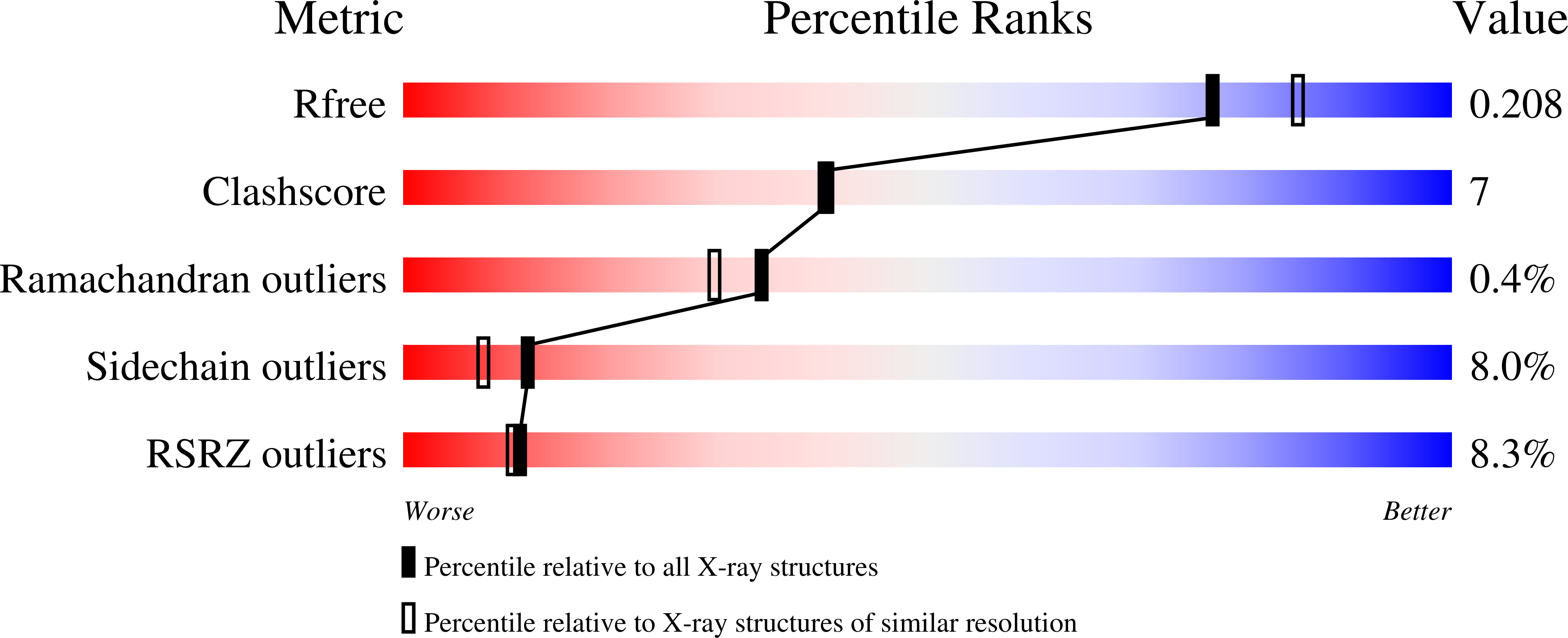
Experimental Data Snapshot
wwPDB Validation 3D Report Full Report
Entity ID: 1 | |||||
|---|---|---|---|---|---|
| Molecule | Chains | Sequence Length | Organism | Details | Image |
| Hypothetical protein VioE | 194 | Chromobacterium violaceum | Mutation(s): 0 | 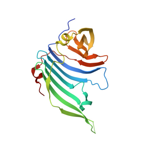 | |
UniProt | |||||
Find proteins for Q7NSZ5 (Chromobacterium violaceum (strain ATCC 12472 / DSM 30191 / JCM 1249 / NBRC 12614 / NCIMB 9131 / NCTC 9757)) Explore Q7NSZ5 Go to UniProtKB: Q7NSZ5 | |||||
Entity Groups | |||||
| Sequence Clusters | 30% Identity50% Identity70% Identity90% Identity95% Identity100% Identity | ||||
| UniProt Group | Q7NSZ5 | ||||
Sequence AnnotationsExpand | |||||
| |||||
| Length ( Å ) | Angle ( ˚ ) |
|---|---|
| a = 84.099 | α = 90 |
| b = 88.435 | β = 90 |
| c = 153.859 | γ = 90 |
| Software Name | Purpose |
|---|---|
| REFMAC | refinement |
| HKL-2000 | data collection |
| HKL-2000 | data reduction |
| HKL-2000 | data scaling |
| MOLREP | phasing |
RCSB PDB (citation) is hosted by
RCSB PDB is a member of the

