2Z8R

Crystal structure of rhamnogalacturonan lyase YesW at 1.40 A resolution


Experimental Data Snapshot

  • Method: X-RAY DIFFRACTION
  • Resolution: 1.40 Å
  • R-Value Free: 0.182 
  • R-Value Work: 0.164 
  • R-Value Observed: 0.164 

wwPDB Validation   3D Report Full Report


This is version 1.2 of the entry. See complete history


Literature

A Novel Structural Fold in Polysaccharide Lyases: BACILLUS SUBTILIS FAMILY 11 RHAMNOGALACTURONAN LYASE YesW WITH AN EIGHT-BLADED -PROPELLER

Ochiai, A.Itoh, T.Maruyama, Y.Kawamata, A.Mikami, B.Hashimoto, W.Murata, K.

(2007) J Biol Chem 282: 37134-37145

  • DOI: https://doi.org/10.1074/jbc.M704663200
  • Primary Citation of Related Structures:  
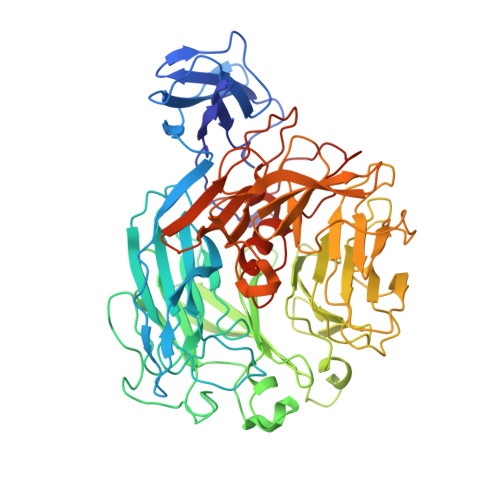
    2Z8R, 2Z8S

  • PubMed Abstract: 

    Rhamnogalacturonan (RG) lyase produced by plant pathogenic and saprophytic microbes plays an important role in degrading plant cell walls. An extracellular RG lyase YesW from saprophytic Bacillus subtilis is a member of polysaccharide lyase family 11 and cleaves glycoside bonds in polygalacturonan as well as RG type-I through a beta-elimination reaction. Crystal structures of YesW and its complex with galacturonan disaccharide, a reaction product analogue, were determined at 1.4 and 2.5 A resolutions with final R-factors of 16.4% and 16.6%, respectively. The enzyme is composed of an eight-bladed beta-propeller with a deep cleft in the center as a basic scaffold, and its structural fold has not been seen in polysaccharide lyases analyzed thus far. Structural analysis of the disaccharide-bound YesW and a site-directed mutagenesis study suggested that Arg-452 and Lys-535 stabilize the carboxyl group of the acidic polysaccharide molecule and Tyr-595 makes a stack interaction with the sugar pyranose ring. In addition to amino acid residues binding to the disaccharide, one calcium ion, which is coordinated by Asp-401, Glu-422, His-363, and His-399, may mediate the enzyme activity. This is, to our knowledge, the first report of a new structural category with a beta-propeller fold in polysaccharide lyases and provides structural insights into substrate binding by RG lyase.


  • Organizational Affiliation

    Laboratory of Basic and Applied Molecular Biotechnology, Graduate School of Agriculture, Kyoto University, Japan.


Macromolecules
Find similar proteins by:  (by identity cutoff)  |  3D Structure
Entity ID: 1
MoleculeChains Sequence LengthOrganismDetailsImage
YesW protein
A, B
591Bacillus subtilisMutation(s): 0 
Gene Names: yesW
EC: 4.2.2
UniProt
Find proteins for O31526 (Bacillus subtilis (strain 168))
Explore O31526 
Go to UniProtKB:  O31526
Entity Groups  
Sequence Clusters30% Identity50% Identity70% Identity90% Identity95% Identity100% Identity
UniProt GroupO31526
Sequence Annotations
Expand
  • Reference Sequence
Small Molecules
Ligands 2 Unique
IDChains Name / Formula / InChI Key2D Diagram3D Interactions
MPD
Query on MPD

Download Ideal Coordinates CCD File 
CA [auth B]
DA [auth B]
EA [auth B]
FA [auth B]
M [auth A]
CA [auth B],
DA [auth B],
EA [auth B],
FA [auth B],
M [auth A],
N [auth A],
O [auth A],
P [auth A],
Q [auth A],
R [auth A]
(4S)-2-METHYL-2,4-PENTANEDIOL
C6 H14 O2
SVTBMSDMJJWYQN-YFKPBYRVSA-N
CA
Query on CA

Download Ideal Coordinates CCD File 
AA [auth B]
BA [auth B]
C [auth A]
D [auth A]
E [auth A]
AA [auth B],
BA [auth B],
C [auth A],
D [auth A],
E [auth A],
F [auth A],
G [auth A],
H [auth A],
I [auth A],
J [auth A],
K [auth A],
L [auth A],
S [auth B],
T [auth B],
U [auth B],
V [auth B],
W [auth B],
X [auth B],
Y [auth B],
Z [auth B]
CALCIUM ION
Ca
BHPQYMZQTOCNFJ-UHFFFAOYSA-N
Experimental Data & Validation

Experimental Data

  • Method: X-RAY DIFFRACTION
  • Resolution: 1.40 Å
  • R-Value Free: 0.182 
  • R-Value Work: 0.164 
  • R-Value Observed: 0.164 
  • Space Group: P 1 21 1
Unit Cell:
Length ( Å )Angle ( ˚ )
a = 56.897α = 90
b = 106.153β = 95.11
c = 101.041γ = 90
Software Package:
Software NamePurpose
REFMACrefinement
HKL-2000data collection
HKL-2000data reduction
HKL-2000data scaling
SOLVEphasing

Structure Validation

View Full Validation Report


Entry History 

Deposition Data

Revision History  (Full details and data files)

  • Version 1.0: 2007-10-16
    Type: Initial release
  • Version 1.1: 2011-07-13
    Changes: Version format compliance
  • Version 1.2: 2024-03-13
    Changes: Data collection, Database references, Derived calculations