2YK3

CRITHIDIA FASCICULATA CYTOCHROME C


Experimental Data Snapshot

  • Method: X-RAY DIFFRACTION
  • Resolution: 1.55 Å
  • R-Value Free: 0.241 
  • R-Value Work: 0.206 
  • R-Value Observed: 0.208 

wwPDB Validation   3D Report Full Report


Ligand Structure Quality Assessment 


This is version 2.1 of the entry. See complete history


Literature

Structure of a Trypanosomatid Mitochondrial Cytochrome C with Heme Attached Via Only One Thioether Bond and Implications for the Substrate Recognition Requirements of Heme Lyase.

Fulop, V.Sam, K.A.Ferguson, S.J.Ginger, M.L.Allen, J.W.A.

(2009) FEBS J 276: 2822

  • DOI: https://doi.org/10.1111/j.1742-4658.2009.07005.x
  • Primary Citation of Related Structures:  
    2YK3

  • PubMed Abstract: 

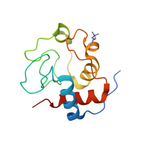
    The principal physiological role of mitochondrial cytochrome c is electron transfer during oxidative phosphorylation. c-Type cytochromes are almost always characterized by covalent attachment of heme to protein through two thioether bonds between the heme vinyl groups and the thiols of cysteine residues in a Cys-Xxx-Xxx-Cys-His motif. Uniquely, however, members of the evolutionarily divergent protist phylum Euglenozoa, which includes Trypanosoma and Leishmania species, have mitochondrial cytochromes c with heme attached through only one thioether bond [to an (A/F)XXCH motif]; the implications of this for the cytochrome structures are unclear. Here we present the 1.55 A resolution X-ray crystal structure of cytochrome c from the trypanosomatid Crithidia fasciculata. Despite the fundamental difference in heme attachment and in the cytochrome c biogenesis machinery of the Euglenozoa, the structure is remarkably similar to that of typical (CXXCH) mitochondrial cytochromes c, both in overall fold and, other than the missing thioether bond, in the details of the heme attachment. Notably, this similarity includes the stereochemistry of the covalent heme attachment to the protein. The structure has implications for the maturation of c-type cytochromes in the Euglenozoa; it also hints at a distinctive redox environment in the mitochondrial intermembrane space of trypanosomes. Surprisingly, Saccharomyces cerevisiae cytochrome c heme lyase (the yeast cytochrome c biogenesis system) cannot efficiently mature Trypanosoma brucei cytochrome c or a CXXCH variant when expressed in the cytoplasm of Escherichia coli, despite their great structural similarity to yeast cytochrome c, suggesting that heme lyase requires specific recognition features in the apocytochrome.


  • Organizational Affiliation

    Department of Biological Sciences, University of Warwick, Coventry, UK.


Macromolecules
Find similar proteins by:  (by identity cutoff)  |  3D Structure
Entity ID: 1
MoleculeChains Sequence LengthOrganismDetailsImage
CYTOCHROME C
A, B, C
114Crithidia fasciculataMutation(s): 0 
UniProt
Find proteins for P00078 (Crithidia fasciculata)
Explore P00078 
Go to UniProtKB:  P00078
Entity Groups  
Sequence Clusters30% Identity50% Identity70% Identity90% Identity95% Identity100% Identity
UniProt GroupP00078
Sequence Annotations
Expand
  • Reference Sequence
Small Molecules
Ligands 2 Unique
IDChains Name / Formula / InChI Key2D Diagram3D Interactions
HEC
Query on HEC

Download Ideal Coordinates CCD File 
D [auth A],
H [auth B],
K [auth C]
HEME C
C34 H34 Fe N4 O4
HXQIYSLZKNYNMH-LJNAALQVSA-N
SO4
Query on SO4

Download Ideal Coordinates CCD File 
E [auth A]
F [auth A]
G [auth A]
I [auth B]
J [auth B]
E [auth A],
F [auth A],
G [auth A],
I [auth B],
J [auth B],
L [auth C],
M [auth C]
SULFATE ION
O4 S
QAOWNCQODCNURD-UHFFFAOYSA-L
Modified Residues  1 Unique
IDChains TypeFormula2D DiagramParent
M3L
Query on M3L
A, B, C
L-PEPTIDE LINKINGC9 H21 N2 O2LYS
Experimental Data & Validation

Experimental Data

  • Method: X-RAY DIFFRACTION
  • Resolution: 1.55 Å
  • R-Value Free: 0.241 
  • R-Value Work: 0.206 
  • R-Value Observed: 0.208 
  • Space Group: C 1 2 1
Unit Cell:
Length ( Å )Angle ( ˚ )
a = 85.93α = 90
b = 110.88β = 131.2
c = 60.93γ = 90
Software Package:
Software NamePurpose
REFMACrefinement
DENZOdata reduction
SCALEPACKdata scaling
PHASERphasing

Structure Validation

View Full Validation Report


Ligand Structure Quality Assessment 


Entry History 

Deposition Data

Revision History  (Full details and data files)

  • Version 1.0: 2011-06-15
    Type: Initial release
  • Version 1.1: 2013-04-17
    Changes: Database references, Version format compliance
  • Version 2.0: 2019-09-25
    Changes: Atomic model, Data collection, Derived calculations, Experimental preparation, Non-polymer description, Other, Structure summary
  • Version 2.1: 2023-12-20
    Changes: Data collection, Database references, Derived calculations, Refinement description