2YFR

Crystal structure of inulosucrase from Lactobacillus johnsonii NCC533


Experimental Data Snapshot

  • Method: X-RAY DIFFRACTION
  • Resolution: 1.75 Å
  • R-Value Free: 0.170 
  • R-Value Work: 0.151 
  • R-Value Observed: 0.152 

wwPDB Validation   3D Report Full Report


This is version 1.2 of the entry. See complete history


Literature

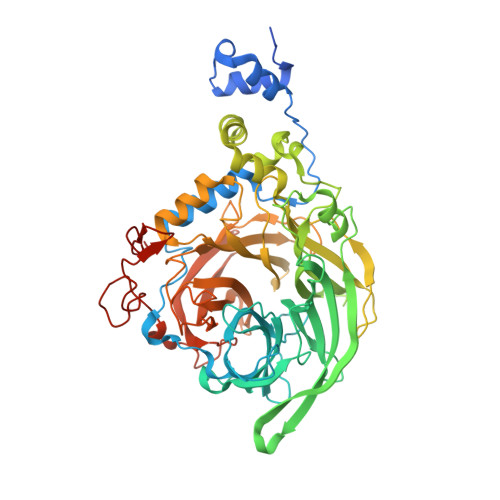
Crystal Structure of Inulosucrase from Lactobacillus: Insights Into the Substrate Specificity and Product Specificity of Gh68 Fructansucrases.

Pijning, T.Anwar, M.A.Boger, M.Dobruchowska, J.M.Leemhuis, H.Kralj, S.Dijkhuizen, L.Dijkstra, B.W.

(2011) J Mol Biol 412: 80

  • DOI: https://doi.org/10.1016/j.jmb.2011.07.031
  • Primary Citation of Related Structures:  
    2YFR, 2YFS, 2YFT

  • PubMed Abstract: 

    Fructansucrases (FSs) catalyze a transfructosylation reaction with sucrose as substrate to produce fructo-oligosaccharides and fructan polymers that contain either β-2,1 glycosidic linkages (inulin) or β-2,6 linkages (levan). Levan-synthesizing FSs (levansucrases) have been most extensively investigated, while detailed information on inulosucrases is limited. Importantly, the molecular basis of the different product specificities of levansucrases and inulosucrases is poorly understood. We have elucidated the three-dimensional structure of a truncated active bacterial GH68 inulosucrase, InuJ of Lactobacillus johnsonii NCC533 (residues 145-708), in its apo form, with a bound substrate (sucrose), and with a transfructosylation product. The sucrose binding pocket and the sucrose binding mode are virtually identical with those of GH68 levansucrases, confirming that both enzyme types use the same fully conserved structural framework for the binding and cleavage of the donor substrate sucrose in the active site. The binding mode of the first transfructosylation product 1-kestose (Fru-β(2-1)-Fru-α(2-1)-Glc, where Fru=fructose and Glc=glucose) in subsites -1 to +2 shows for the first time how inulin-type fructo-oligosaccharide bind in GH68 FS and how an inulin-type linkage can be formed. Surprisingly, observed interactions with the sugar in subsites +1 and +2 are provided by residues that are also present in levansucrases. The binding mode of 1-kestose and the presence of a more distant sucrose binding site suggest that residues beyond the +2 subsite, in particular residues from the nonconserved 1B-1C loop, determine product linkage type specificity in GH68 FSs.


  • Organizational Affiliation

    Laboratory of Biophysical Chemistry, Groningen Biomolecular Sciences and Biotechnology Institute, University of Groningen, Nijenborgh 7, 9747 AG Groningen, The Netherlands.


Macromolecules
Find similar proteins by:  (by identity cutoff)  |  3D Structure
Entity ID: 1
MoleculeChains Sequence LengthOrganismDetailsImage
LEVANSUCRASE571Lactobacillus johnsoniiMutation(s): 0 
EC: 2.4.1.9
UniProt
Find proteins for Q74K42 (Lactobacillus johnsonii (strain CNCM I-12250 / La1 / NCC 533))
Explore Q74K42 
Go to UniProtKB:  Q74K42
Entity Groups  
Sequence Clusters30% Identity50% Identity70% Identity90% Identity95% Identity100% Identity
UniProt GroupQ74K42
Sequence Annotations
Expand
  • Reference Sequence
Small Molecules
Ligands 5 Unique
IDChains Name / Formula / InChI Key2D Diagram3D Interactions
SO4
Query on SO4

Download Ideal Coordinates CCD File 
G [auth A]
H [auth A]
I [auth A]
J [auth A]
K [auth A]
G [auth A],
H [auth A],
I [auth A],
J [auth A],
K [auth A],
L [auth A],
M [auth A],
N [auth A],
O [auth A]
SULFATE ION
O4 S
QAOWNCQODCNURD-UHFFFAOYSA-L
GOL
Query on GOL

Download Ideal Coordinates CCD File 
P [auth A]
Q [auth A]
R [auth A]
S [auth A]
T [auth A]
P [auth A],
Q [auth A],
R [auth A],
S [auth A],
T [auth A],
U [auth A],
V [auth A],
W [auth A],
X [auth A]
GLYCEROL
C3 H8 O3
PEDCQBHIVMGVHV-UHFFFAOYSA-N
ACT
Query on ACT

Download Ideal Coordinates CCD File 
D [auth A],
E [auth A],
F [auth A]
ACETATE ION
C2 H3 O2
QTBSBXVTEAMEQO-UHFFFAOYSA-M
CA
Query on CA

Download Ideal Coordinates CCD File 
B [auth A]CALCIUM ION
Ca
BHPQYMZQTOCNFJ-UHFFFAOYSA-N
CL
Query on CL

Download Ideal Coordinates CCD File 
C [auth A]CHLORIDE ION
Cl
VEXZGXHMUGYJMC-UHFFFAOYSA-M
Experimental Data & Validation

Experimental Data

  • Method: X-RAY DIFFRACTION
  • Resolution: 1.75 Å
  • R-Value Free: 0.170 
  • R-Value Work: 0.151 
  • R-Value Observed: 0.152 
  • Space Group: I 4 2 2
Unit Cell:
Length ( Å )Angle ( ˚ )
a = 172.007α = 90
b = 172.007β = 90
c = 114.581γ = 90
Software Package:
Software NamePurpose
REFMACrefinement
XDSdata reduction
XDSdata scaling
PHASERphasing

Structure Validation

View Full Validation Report


Entry History 

Deposition Data

Revision History  (Full details and data files)

  • Version 1.0: 2011-08-03
    Type: Initial release
  • Version 1.1: 2011-08-31
    Changes: Database references
  • Version 1.2: 2023-12-20
    Changes: Data collection, Database references, Derived calculations, Other, Refinement description