2YAU

X-ray structure of the Leishmania infantum tryopanothione reductase in complex with auranofin


Experimental Data Snapshot

  • Method: X-RAY DIFFRACTION
  • Resolution: 3.50 Å
  • R-Value Free: 0.319 
  • R-Value Work: 0.267 
  • R-Value Observed: 0.270 

wwPDB Validation   3D Report Full Report


Ligand Structure Quality Assessment 


This is version 1.3 of the entry. See complete history


Literature

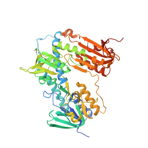
A Gold-Containing Drug Against Parasitic Polyamine Metabolism: The X-Ray Structure of Trypanothione Reductase from Leishmania Infantum in Complex with Auranofin Reveals a Dual Mechanism of Enzyme Inhibition.

Ilari, A.Baiocco, P.Messori, L.Fiorillo, A.Boffi, A.Gramiccia, M.Di Muccio, T.Colotti, G.

(2012) Amino Acids 42: 803

  • DOI: https://doi.org/10.1007/s00726-011-0997-9
  • Primary Citation of Related Structures:  
    2YAU

  • PubMed Abstract: 

    Auranofin is a gold(I)-containing drug in clinical use as an antiarthritic agent. Recent studies showed that auranofin manifests interesting antiparasitic actions very likely arising from inhibition of parasitic enzymes involved in the control of the redox metabolism. Trypanothione reductase is a key enzyme of Leishmania infantum polyamine-dependent redox metabolism, and a validated target for antileishmanial drugs. As trypanothione reductase contains a dithiol motif at its active site and gold(I) compounds are known to be highly thiophilic, we explored whether auranofin might behave as an effective enzyme inhibitor and as a potential antileishmanial agent. Notably, enzymatic assays revealed that auranofin causes indeed a pronounced enzyme inhibition. To gain a deeper insight into the molecular basis of enzyme inhibition, crystals of the auranofin-bound enzyme, in the presence of NADPH, were prepared, and the X-ray crystal structure of the auranofin-trypanothione reductase-NADPH complex was solved at 3.5 Å resolution. In spite of the rather low resolution, these data were of sufficient quality as to identify the presence of the gold center and of the thiosugar of auranofin, and to locate them within the overall protein structure. Gold binds to the two active site cysteine residues of TR, i.e. Cys52 and Cys57, while the thiosugar moiety of auranofin binds to the trypanothione binding site; thus auranofin appears to inhibit TR through a dual mechanism. Auranofin kills the promastigote stage of L. infantum at micromolar concentration; these findings will contribute to the design of new drugs against leishmaniasis.


  • Organizational Affiliation

    Department of Biochemical Sciences, Institute of Molecular Biology and Pathology CNR, Sapienza University of Rome, P.le A. Moro 5, 00185, Rome, Italy. Andrea.ilari@uniroma1.it


Macromolecules
Find similar proteins by:  (by identity cutoff)  |  3D Structure
Entity ID: 1
MoleculeChains Sequence LengthOrganismDetailsImage
TRYPANOTHIONE REDUCTASE
A, B
511Leishmania infantumMutation(s): 0 
EC: 1.8.1.12
UniProt
Find proteins for A4HSF7 (Leishmania infantum)
Explore A4HSF7 
Go to UniProtKB:  A4HSF7
Entity Groups  
Sequence Clusters30% Identity50% Identity70% Identity90% Identity95% Identity100% Identity
UniProt GroupA4HSF7
Sequence Annotations
Expand
  • Reference Sequence
Small Molecules
Ligands 6 Unique
IDChains Name / Formula / InChI Key2D Diagram3D Interactions
FAD
Query on FAD

Download Ideal Coordinates CCD File 
C [auth A],
I [auth B]
FLAVIN-ADENINE DINUCLEOTIDE
C27 H33 N9 O15 P2
VWWQXMAJTJZDQX-UYBVJOGSSA-N
NDP
Query on NDP

Download Ideal Coordinates CCD File 
D [auth A],
J [auth B]
NADPH DIHYDRO-NICOTINAMIDE-ADENINE-DINUCLEOTIDE PHOSPHATE
C21 H30 N7 O17 P3
ACFIXJIJDZMPPO-NNYOXOHSSA-N
TS8
Query on TS8

Download Ideal Coordinates CCD File 
F [auth A],
L [auth B]
2,3,4,6-tetra-O-acetyl-1-thio-beta-D-glucopyranose
C14 H20 O9 S
SFOZKJGZNOBSHF-RGDJUOJXSA-N
AU
Query on AU

Download Ideal Coordinates CCD File 
E [auth A],
K [auth B]
GOLD ION
Au
ZBKIUFWVEIBQRT-UHFFFAOYSA-N
SO4
Query on SO4

Download Ideal Coordinates CCD File 
H [auth A],
N [auth B]
SULFATE ION
O4 S
QAOWNCQODCNURD-UHFFFAOYSA-L
CL
Query on CL

Download Ideal Coordinates CCD File 
G [auth A],
M [auth B]
CHLORIDE ION
Cl
VEXZGXHMUGYJMC-UHFFFAOYSA-M
Experimental Data & Validation

Experimental Data

  • Method: X-RAY DIFFRACTION
  • Resolution: 3.50 Å
  • R-Value Free: 0.319 
  • R-Value Work: 0.267 
  • R-Value Observed: 0.270 
  • Space Group: P 41
Unit Cell:
Length ( Å )Angle ( ˚ )
a = 103.058α = 90
b = 103.058β = 90
c = 191.604γ = 90
Software Package:
Software NamePurpose
REFMACrefinement
DENZOdata reduction
SCALEPACKdata scaling
MOLREPphasing

Structure Validation

View Full Validation Report


Ligand Structure Quality Assessment 


Entry History 

Deposition Data

Revision History  (Full details and data files)

  • Version 1.0: 2012-01-11
    Type: Initial release
  • Version 1.1: 2012-02-08
    Changes: Other
  • Version 1.2: 2020-07-29
    Type: Remediation
    Reason: Carbohydrate remediation
    Changes: Data collection, Derived calculations, Other, Structure summary
  • Version 1.3: 2023-12-20
    Changes: Data collection, Database references, Refinement description, Structure summary