2Y67

New 5-Benzylidenethiazolidine-4-one Inhibitors of Bacterial MurD Ligase: Design, Synthesis, Crystal Structures, and Biological Evaluation


Experimental Data Snapshot

  • Method: X-RAY DIFFRACTION
  • Resolution: 1.85 Å
  • R-Value Free: 0.249 
  • R-Value Work: 0.208 
  • R-Value Observed: 0.212 

wwPDB Validation   3D Report Full Report


Ligand Structure Quality Assessment 


This is version 1.2 of the entry. See complete history


Literature

New 5-Benzylidenethiazolidin-4-One Inhibitors of Bacterial Murd Ligase: Design, Synthesis, Crystal Structures, and Biological Evaluation.

Zidar, N.Tomasic, T.Sink, R.Kovac, A.Patin, D.Blanot, D.Contreras-Martel, C.Dessen, A.Premru, M.M.Zega, A.Gobec, S.Masic, L.P.Kikelj, D.

(2011) Eur J Med Chem 46: 5512

  • DOI: https://doi.org/10.1016/j.ejmech.2011.09.017
  • Primary Citation of Related Structures:  
    2Y66, 2Y67

  • PubMed Abstract: 

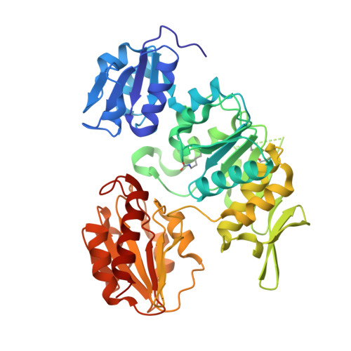
    Mur ligases (MurC-MurF), a group of bacterial enzymes that catalyze four consecutive steps in the formation of cytoplasmic peptidoglycan precursor, are becoming increasingly adopted as targets in antibacterial drug design. Based on the crystal structure of MurD cocrystallized with thiazolidine-2,4-dione inhibitor I, we have designed, synthesized, and evaluated a series of improved glutamic acid containing 5-benzylidenerhodanine and 5-benzylidenethiazolidine-2,4-dione inhibitors of MurD with IC(50) values up to 28 μM. Inhibitor 37, with an IC(50) of 34 μM, displays a weak antibacterial activity against S. aureus ATCC 29213 and E. faecalis ATCC 29212 with minimal inhibitory concentrations of 128 μg/mL. High-resolution crystal structures of MurD in complex with two new inhibitors (compounds 23 and 51) reveal details of their binding modes within the active site and provide valuable information for further structure-based optimization.


  • Organizational Affiliation

    Faculty of Pharmacy, University of Ljubljana, 1000 Ljubljana, Slovenia.


Macromolecules
Find similar proteins by:  (by identity cutoff)  |  3D Structure
Entity ID: 1
MoleculeChains Sequence LengthOrganismDetailsImage
UDP-N-ACETYLMURAMOYLALANINE--D-GLUTAMATE LIGASE445Escherichia coli K-12Mutation(s): 0 
EC: 6.3.2.9
UniProt
Find proteins for P14900 (Escherichia coli (strain K12))
Explore P14900 
Go to UniProtKB:  P14900
Entity Groups  
Sequence Clusters30% Identity50% Identity70% Identity90% Identity95% Identity100% Identity
UniProt GroupP14900
Sequence Annotations
Expand
  • Reference Sequence
Small Molecules
Ligands 2 Unique
IDChains Name / Formula / InChI Key2D Diagram3D Interactions
N21
Query on N21

Download Ideal Coordinates CCD File 
B [auth A](2R)-2-[[4-[[4-[(Z)-(2,4-dioxo-1,3-thiazolidin-5-ylidene)methyl]phenoxy]methyl]phenyl]sulfonylamino]pentanedioic acid
C22 H20 N2 O9 S2
BZCXCLVFYJNKLW-LVSMMTLPSA-N
SO4
Query on SO4

Download Ideal Coordinates CCD File 
C [auth A]SULFATE ION
O4 S
QAOWNCQODCNURD-UHFFFAOYSA-L
Modified Residues  1 Unique
IDChains TypeFormula2D DiagramParent
KCX
Query on KCX
A
L-PEPTIDE LINKINGC7 H14 N2 O4LYS
Binding Affinity Annotations 
IDSourceBinding Affinity
N21 Binding MOAD:  2Y67 IC50: 2.92e+5 (nM) from 1 assay(s)
PDBBind:  2Y67 IC50: 2.92e+5 (nM) from 1 assay(s)
Experimental Data & Validation

Experimental Data

  • Method: X-RAY DIFFRACTION
  • Resolution: 1.85 Å
  • R-Value Free: 0.249 
  • R-Value Work: 0.208 
  • R-Value Observed: 0.212 
  • Space Group: P 41
Unit Cell:
Length ( Å )Angle ( ˚ )
a = 65.326α = 90
b = 65.326β = 90
c = 134.707γ = 90
Software Package:
Software NamePurpose
REFMACrefinement
XDSdata reduction
XSCALEdata scaling
PHASERphasing

Structure Validation

View Full Validation Report


Ligand Structure Quality Assessment 


Entry History 

Revision History  (Full details and data files)

  • Version 1.0: 2011-10-19
    Type: Initial release
  • Version 1.1: 2011-11-02
    Changes: Database references
  • Version 1.2: 2023-12-20
    Changes: Data collection, Database references, Derived calculations, Other, Refinement description