2Y0N

CRYSTAL STRUCTURE OF THE COMPLEX BETWEEN DOSAGE COMPENSATION FACTORS MSL1 AND MSL3


Experimental Data Snapshot

  • Method: X-RAY DIFFRACTION
  • Resolution: 3.00 Å
  • R-Value Free: 0.253 
  • R-Value Work: 0.232 
  • R-Value Observed: 0.233 

wwPDB Validation   3D Report Full Report


This is version 1.3 of the entry. See complete history


Literature

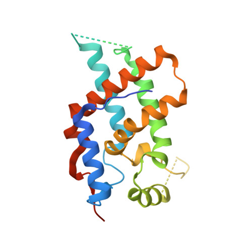
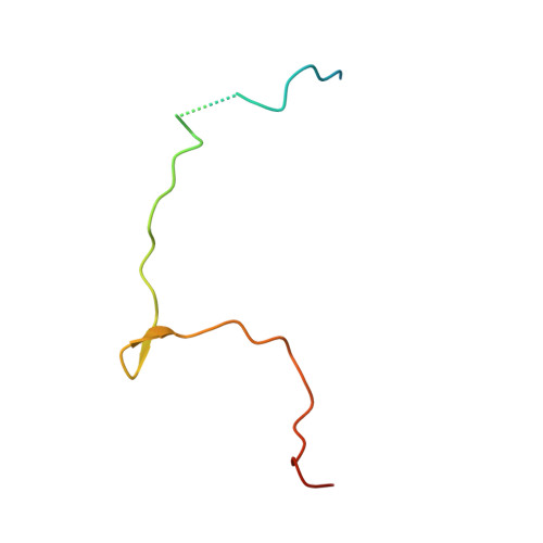
Structural Basis for Mof and Msl3 Recruitment Into the Dosage Compensation Complex by Msl1.

Kadlec, J.Hallacli, E.Lipp, M.Holz, H.Sanchez-Weatherby, J.Cusack, S.Akhtar, A.

(2011) Nat Struct Mol Biol 18: 142

  • DOI: https://doi.org/10.1038/nsmb.1960
  • Primary Citation of Related Structures:  
    2Y0M, 2Y0N

  • PubMed Abstract: 

    The male-specific lethal (MSL) complex is required for dosage compensation in Drosophila melanogaster, and analogous complexes exist in mammals. We report structures of binary complexes of mammalian MSL3 and the histone acetyltransferase (HAT) MOF with consecutive segments of MSL1. MSL1 interacts with MSL3 as an extended chain forming an extensive hydrophobic interface, whereas the MSL1-MOF interface involves electrostatic interactions between the HAT domain and a long helix of MSL1. This structure provides insights into the catalytic mechanism of MOF and enables us to show analogous interactions of MOF with NSL1. In Drosophila, selective disruption of Msl1 interactions with Msl3 or Mof severely affects Msl1 targeting to the body of dosage-compensated genes and several high-affinity sites, without affecting promoter binding. We propose that Msl1 acts as a scaffold for MSL complex assembly to achieve specific targeting to the X chromosome.


  • Organizational Affiliation

    European Molecular Biology Laboratory, Grenoble, France.


Macromolecules
Find similar proteins by:  (by identity cutoff)  |  3D Structure
Entity ID: 1
MoleculeChains Sequence LengthOrganismDetailsImage
MALE-SPECIFIC LETHAL 3 HOMOLOG
A, B, C, D
211Homo sapiensMutation(s): 0 
UniProt & NIH Common Fund Data Resources
Find proteins for Q8N5Y2 (Homo sapiens)
Explore Q8N5Y2 
Go to UniProtKB:  Q8N5Y2
PHAROS:  Q8N5Y2
GTEx:  ENSG00000005302 
Entity Groups  
Sequence Clusters30% Identity50% Identity70% Identity90% Identity95% Identity100% Identity
UniProt GroupQ8N5Y2
Sequence Annotations
Expand
  • Reference Sequence
Find similar proteins by:  (by identity cutoff)  |  3D Structure
Entity ID: 2
MoleculeChains Sequence LengthOrganismDetailsImage
MALE-SPECIFIC LETHAL 1 HOMOLOG
E, F, G, H
56Mus musculusMutation(s): 0 
UniProt & NIH Common Fund Data Resources
Find proteins for Q6PDM1 (Mus musculus)
Explore Q6PDM1 
Go to UniProtKB:  Q6PDM1
IMPC:  MGI:1921276
Entity Groups  
Sequence Clusters30% Identity50% Identity70% Identity90% Identity95% Identity100% Identity
UniProt GroupQ6PDM1
Sequence Annotations
Expand
  • Reference Sequence
Experimental Data & Validation

Experimental Data

  • Method: X-RAY DIFFRACTION
  • Resolution: 3.00 Å
  • R-Value Free: 0.253 
  • R-Value Work: 0.232 
  • R-Value Observed: 0.233 
  • Space Group: P 1 21 1
Unit Cell:
Length ( Å )Angle ( ˚ )
a = 76.23α = 90
b = 127.27β = 118.46
c = 79.68γ = 90
Software Package:
Software NamePurpose
REFMACrefinement
XDSdata reduction
XSCALEdata scaling
PHASERphasing

Structure Validation

View Full Validation Report


Entry History 

Deposition Data

Revision History  (Full details and data files)

  • Version 1.0: 2011-01-12
    Type: Initial release
  • Version 1.1: 2011-05-08
    Changes: Version format compliance
  • Version 1.2: 2011-07-13
    Changes: Version format compliance
  • Version 1.3: 2023-12-20
    Changes: Data collection, Database references, Other, Refinement description