2XVG

crystal structure of alpha-xylosidase (GH31) from Cellvibrio japonicus


Experimental Data Snapshot

  • Method: X-RAY DIFFRACTION
  • Resolution: 2.60 Å
  • R-Value Free: 0.259 
  • R-Value Work: 0.204 
  • R-Value Observed: 0.206 

wwPDB Validation   3D Report Full Report


This is version 1.3 of the entry. See complete history


Literature

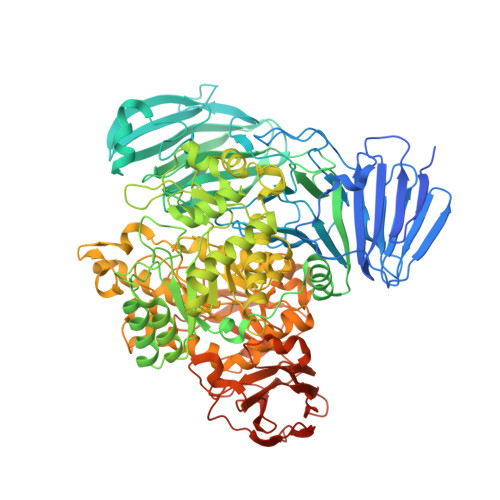
Structural and Enzymatic Characterisation of a Glycoside Hydrolase Family 31 Alpha-Xylosidase from Cellvibrio Japonicus Involved in Xyloglucan Saccharification.

Larsbrink, J.Izumi, A.Ibatullin, F.Nakhai, A.Gilbert, H.J.Davies, G.J.Brumer, H.

(2011) Biochem J 436: 567

  • DOI: https://doi.org/10.1042/BJ20110299
  • Primary Citation of Related Structures:  
    2XVG, 2XVK, 2XVL

  • PubMed Abstract: 

    The desire for improved methods of biomass conversion into fuels and feedstocks has re-awakened interest in the enzymology of plant cell wall degradation. The complex polysaccharide xyloglucan is abundant in plant matter, where it may account for up to 20% of the total primary cell wall carbohydrates. Despite this, few studies have focused on xyloglucan saccharification, which requires a consortium of enzymes including endo-xyloglucanases, α-xylosidases, β-galactosidases and α-L-fucosidases, among others. In the present paper, we show the characterization of Xyl31A, a key α-xylosidase in xyloglucan utilization by the model Gram-negative soil saprophyte Cellvibrio japonicus. CjXyl31A exhibits high regiospecificity for the hydrolysis of XGOs (xylogluco-oligosaccharides), with a particular preference for longer substrates. Crystallographic structures of both the apo enzyme and the trapped covalent 5-fluoro-β-xylosyl-enzyme intermediate, together with docking studies with the XXXG heptasaccharide, revealed, for the first time in GH31 (glycoside hydrolase family 31), the importance of a PA14 domain insert in the recognition of longer oligosaccharides by extension of the active-site pocket. The observation that CjXyl31A was localized to the outer membrane provided support for a biological model of xyloglucan utilization by C. japonicus, in which XGOs generated by the action of a secreted endo-xyloglucanase are ultimately degraded in close proximity to the cell surface. Moreover, the present study diversifies the toolbox of glycosidases for the specific modification and saccharification of cell wall polymers for biotechnological applications.


  • Organizational Affiliation

    Division of Glycoscience, School of Biotechnology, Royal Institute of Technology (KTH), AlbaNova University Centre, 106 91 Stockholm, Sweden.


Macromolecules
Find similar proteins by:  (by identity cutoff)  |  3D Structure
Entity ID: 1
MoleculeChains Sequence LengthOrganismDetailsImage
ALPHA XYLOSIDASE1,020Cellvibrio japonicusMutation(s): 0 
EC: 3.2.1
UniProt
Find proteins for B3PBD9 (Cellvibrio japonicus (strain Ueda107))
Explore B3PBD9 
Go to UniProtKB:  B3PBD9
Entity Groups  
Sequence Clusters30% Identity50% Identity70% Identity90% Identity95% Identity100% Identity
UniProt GroupB3PBD9
Sequence Annotations
Expand
  • Reference Sequence
Small Molecules
Ligands 4 Unique
IDChains Name / Formula / InChI Key2D Diagram3D Interactions
SO4
Query on SO4

Download Ideal Coordinates CCD File 
G [auth A]SULFATE ION
O4 S
QAOWNCQODCNURD-UHFFFAOYSA-L
EDO
Query on EDO

Download Ideal Coordinates CCD File 
F [auth A],
H [auth A],
I [auth A]
1,2-ETHANEDIOL
C2 H6 O2
LYCAIKOWRPUZTN-UHFFFAOYSA-N
NI
Query on NI

Download Ideal Coordinates CCD File 
J [auth A],
K [auth A]
NICKEL (II) ION
Ni
VEQPNABPJHWNSG-UHFFFAOYSA-N
CL
Query on CL

Download Ideal Coordinates CCD File 
B [auth A],
C [auth A],
D [auth A],
E [auth A]
CHLORIDE ION
Cl
VEXZGXHMUGYJMC-UHFFFAOYSA-M
Experimental Data & Validation

Experimental Data

  • Method: X-RAY DIFFRACTION
  • Resolution: 2.60 Å
  • R-Value Free: 0.259 
  • R-Value Work: 0.204 
  • R-Value Observed: 0.206 
  • Space Group: P 63 2 2
Unit Cell:
Length ( Å )Angle ( ˚ )
a = 156.193α = 90
b = 156.193β = 90
c = 227.759γ = 120
Software Package:
Software NamePurpose
PHENIXrefinement
MOSFLMdata reduction
SCALEPACKdata scaling
BALBESphasing

Structure Validation

View Full Validation Report


Entry History 

Deposition Data

Revision History  (Full details and data files)

  • Version 1.0: 2011-04-13
    Type: Initial release
  • Version 1.1: 2011-06-02
    Changes: Version format compliance
  • Version 1.2: 2011-07-13
    Changes: Version format compliance
  • Version 1.3: 2023-12-20
    Changes: Data collection, Database references, Derived calculations, Other, Refinement description