2XJ4

Structure of the bacterial cell division regulator protein MipZ


Experimental Data Snapshot

  • Method: X-RAY DIFFRACTION
  • Resolution: 1.60 Å
  • R-Value Free: 0.175 
  • R-Value Work: 0.126 
  • R-Value Observed: 0.129 

wwPDB Validation   3D Report Full Report


This is version 1.2 of the entry. See complete history


Literature

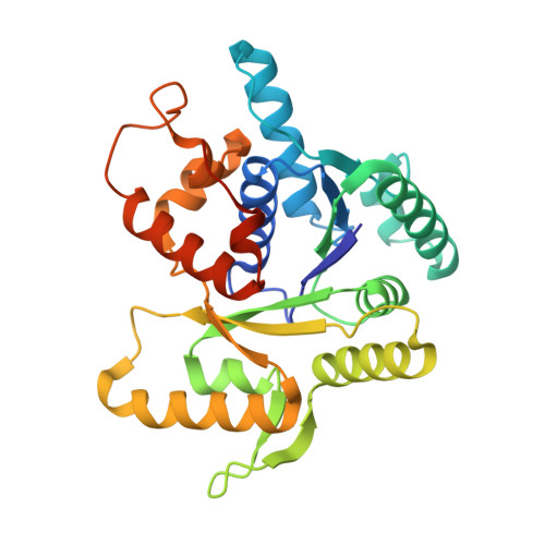
Localized Dimerization and Nucleoid Binding Drive Gradient Formation by the Bacterial Cell Division Inhibitor Mipz.

Kiekebusch, D.Michie, K.A.Essen, L.O.Lowe, J.Thanbichler, M.

(2012) Mol Cell 46: 245

  • DOI: https://doi.org/10.1016/j.molcel.2012.03.004
  • Primary Citation of Related Structures:  
    2XIT, 2XJ4, 2XJ9

  • PubMed Abstract: 

    Protein gradients play a central role in the spatial organization of cells, but the mechanisms of their formation are incompletely understood. This study analyzes the determinants responsible for establishing bipolar gradients of the ATPase MipZ, a key regulator of division site placement in Caulobacter crescentus. We have solved the crystal structure of MipZ in different nucleotide states, dissected its ATPase cycle, and investigated its interaction with FtsZ, ParB, and the nucleoid. Our results suggest that the polar ParB complexes locally stimulate the formation of ATP-bound MipZ dimers, which are then retained near the cell poles through association with chromosomal DNA. Due to their intrinsic ATPase activity, dimers eventually dissociate into freely diffusible monomers that undergo spontaneous nucleotide exchange and are recaptured by ParB. These findings clarify the molecular function of a conserved gradient-forming system and reveal mechanistic principles that might be commonly used to sustain protein gradients within cells.


  • Organizational Affiliation

    Max Planck Institute for Terrestrial Microbiology, 35043 Marburg, Germany.


Macromolecules
Find similar proteins by:  (by identity cutoff)  |  3D Structure
Entity ID: 1
MoleculeChains Sequence LengthOrganismDetailsImage
MIPZ286Caulobacter vibrioidesMutation(s): 0 
UniProt
Find proteins for A0A0H3CA70 (Caulobacter vibrioides (strain NA1000 / CB15N))
Explore A0A0H3CA70 
Go to UniProtKB:  A0A0H3CA70
Entity Groups  
Sequence Clusters30% Identity50% Identity70% Identity90% Identity95% Identity100% Identity
UniProt GroupA0A0H3CA70
Sequence Annotations
Expand
  • Reference Sequence
Experimental Data & Validation

Experimental Data

  • Method: X-RAY DIFFRACTION
  • Resolution: 1.60 Å
  • R-Value Free: 0.175 
  • R-Value Work: 0.126 
  • R-Value Observed: 0.129 
  • Space Group: H 3
Unit Cell:
Length ( Å )Angle ( ˚ )
a = 81.334α = 90
b = 81.334β = 90
c = 124.206γ = 120
Software Package:
Software NamePurpose
REFMACrefinement
SCALAdata scaling
SHELXDphasing

Structure Validation

View Full Validation Report


Entry History 

Deposition Data

Revision History  (Full details and data files)

  • Version 1.0: 2011-07-13
    Type: Initial release
  • Version 1.1: 2012-05-23
    Changes: Database references, Other, Version format compliance
  • Version 1.2: 2012-05-30
    Changes: Other