2XH3

extracellular nuclease


Experimental Data Snapshot

  • Method: X-RAY DIFFRACTION
  • Resolution: 2.49 Å
  • R-Value Free: 0.244 
  • R-Value Work: 0.167 
  • R-Value Observed: 0.171 

wwPDB Validation   3D Report Full Report


This is version 1.3 of the entry. See complete history


Literature

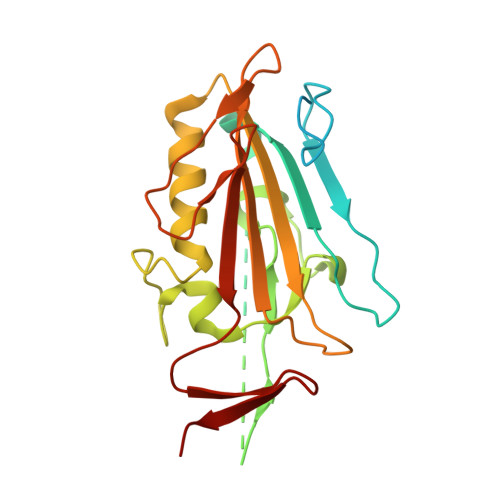
The Structural Characterization of a Prophage-Encoded Extracellular DNase from Streptococcus Pyogenes.

Korczynska, J.E.Turkenburg, J.P.Taylor, E.J.

(2012) Nucleic Acids Res 40: 928

  • DOI: https://doi.org/10.1093/nar/gkr789
  • Primary Citation of Related Structures:  
    2XGR, 2XH3

  • PubMed Abstract: 

    The pathogenic bacterium Group A Streptococcus pyogenes produces several extracellular DNases that have been shown to facilitate invasive infection by evading the human host immune system. DNases degrade the chromatin in neutrophil extracellular traps, enabling the bacterium to evade neutrophil capture. Spd1 is a type I, nonspecific ββα/metal-dependent nuclease from Streptococcus pyogenes, which is encoded by the SF370.1 prophage and is likely to be expressed as a result of prophage induction. We present here the X-ray structure of this DNase in the wild-type and Asn145Ala mutant form. Through structural and sequence alignments as well as mutagenesis studies, we have identified the key residues His121, Asn145 and Glu164, which are crucial for Spd1 nucleolytic activity and shown the active site constellation. Our wild-type structure alludes to the possibility of a catalytically blocked dimeric form of the protein. We have investigated the multimeric nature of Spd1 using size-exclusion chromatography with multi-angle light scattering (SEC-MALLS) in the presence and absence of the divalent metal ion Mg(2+), which suggests that Spd1 exists in a monomeric form in solution.


  • Organizational Affiliation

    Department of Chemistry, Structural Biology Laboratory, The University of York, YO10 5YW, UK.


Macromolecules
Find similar proteins by:  (by identity cutoff)  |  3D Structure
Entity ID: 1
MoleculeChains Sequence LengthOrganismDetailsImage
SPD1 NUCLEASE
A, B
252Streptococcus pyogenes serotype M1Mutation(s): 1 
EC: 3.1.21.1
UniProt
Find proteins for Q9A0M1 (Streptococcus pyogenes serotype M1)
Explore Q9A0M1 
Go to UniProtKB:  Q9A0M1
Entity Groups  
Sequence Clusters30% Identity50% Identity70% Identity90% Identity95% Identity100% Identity
UniProt GroupQ9A0M1
Sequence Annotations
Expand
  • Reference Sequence
Small Molecules
Ligands 1 Unique
IDChains Name / Formula / InChI Key2D Diagram3D Interactions
PEG
Query on PEG

Download Ideal Coordinates CCD File 
C [auth B]DI(HYDROXYETHYL)ETHER
C4 H10 O3
MTHSVFCYNBDYFN-UHFFFAOYSA-N
Experimental Data & Validation

Experimental Data

  • Method: X-RAY DIFFRACTION
  • Resolution: 2.49 Å
  • R-Value Free: 0.244 
  • R-Value Work: 0.167 
  • R-Value Observed: 0.171 
  • Space Group: P 21 21 21
Unit Cell:
Length ( Å )Angle ( ˚ )
a = 49.822α = 90
b = 101.822β = 90
c = 113.547γ = 90
Software Package:
Software NamePurpose
REFMACrefinement
MOSFLMdata reduction
PHASERphasing

Structure Validation

View Full Validation Report


Entry History 

Deposition Data

Revision History  (Full details and data files)

  • Version 1.0: 2011-07-20
    Type: Initial release
  • Version 1.1: 2011-10-19
    Changes: Database references
  • Version 1.2: 2012-01-25
    Changes: Other
  • Version 1.3: 2023-12-20
    Changes: Data collection, Database references, Derived calculations, Other, Refinement description