2XGL

The X-ray structure of the Escherichia coli colicin M immunity protein demonstrates the presence of a disulphide bridge, which is functionally essential


Experimental Data Snapshot

  • Method: X-RAY DIFFRACTION
  • Resolution: 2.70 Å
  • R-Value Free: 0.268 
  • R-Value Work: 0.222 
  • R-Value Observed: 0.224 

wwPDB Validation   3D Report Full Report


This is version 1.3 of the entry. See complete history


Literature

X-Ray Structure and Site-Directed Mutagenesis Analysis of the Escherichia Coli Colicin M Immunity Protein.

Gerard, F.Brooks, M.A.Barreteau, H.Touze, T.Graille, M.Bouhss, A.Blanot, D.Tilbeurgh, H.V.Mengin-Lecreulx, D.

(2011) J Bacteriol 193: 205

  • DOI: https://doi.org/10.1128/JB.01119-10
  • Primary Citation of Related Structures:  
    2XGL

  • PubMed Abstract: 

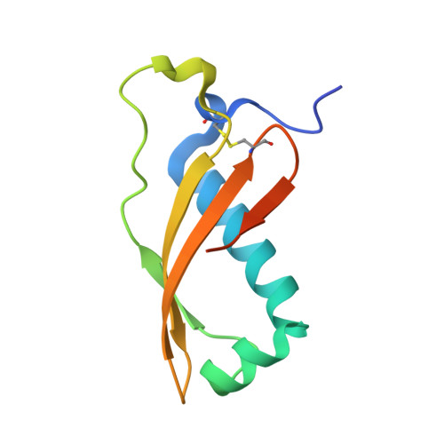
    Colicin M (ColM), which is produced by some Escherichia coli strains to kill competitor strains from the same or related species, was recently shown to inhibit cell wall peptidoglycan biosynthesis through enzymatic degradation of its lipid II precursor. ColM-producing strains are protected from the toxin that they produce by coexpression of a specific immunity protein, named Cmi, whose mode of action still remains to be identified. We report here the resolution of the crystal structure of Cmi, which is composed of four β strands and four α helices. This rather compact structure revealed a disulfide bond between residues Cys31 and Cys107. Interestingly, these two cysteines and several other residues appeared to be conserved in the sequences of several proteins of unknown function belonging to the YebF family which exhibit 25 to 35% overall sequence similarity with Cmi. Site-directed mutagenesis was performed to assess the role of these residues in the ColM immunity-conferring activity of Cmi, which showed that the disulfide bond and residues from the C-terminal extremity of the protein were functionally essential. The involvement of DsbA oxidase in the formation of the Cmi disulfide bond is also demonstrated.


  • Organizational Affiliation

    Laboratoire des Enveloppes Bactériennes et Antibiotiques, Institut de Biochimie et Biophysique Moléculaire et Cellulaire, UMR 8619, Université Paris-Sud, Orsay F-91405, France.


Macromolecules
Find similar proteins by:  (by identity cutoff)  |  3D Structure
Entity ID: 1
MoleculeChains Sequence LengthOrganismDetailsImage
COLICIN-M IMMUNITY PROTEIN
A, B
103Escherichia coliMutation(s): 0 
UniProt
Find proteins for P18002 (Escherichia coli)
Explore P18002 
Go to UniProtKB:  P18002
Entity Groups  
Sequence Clusters30% Identity50% Identity70% Identity90% Identity95% Identity100% Identity
UniProt GroupP18002
Sequence Annotations
Expand
  • Reference Sequence
Small Molecules
Ligands 3 Unique
IDChains Name / Formula / InChI Key2D Diagram3D Interactions
CD
Query on CD

Download Ideal Coordinates CCD File 
C [auth A]
D [auth A]
E [auth A]
F [auth A]
G [auth A]
C [auth A],
D [auth A],
E [auth A],
F [auth A],
G [auth A],
U [auth B],
V [auth B]
CADMIUM ION
Cd
WLZRMCYVCSSEQC-UHFFFAOYSA-N
CL
Query on CL

Download Ideal Coordinates CCD File 
AA [auth B]
BA [auth B]
H [auth A]
L [auth A]
M [auth A]
AA [auth B],
BA [auth B],
H [auth A],
L [auth A],
M [auth A],
O [auth A],
P [auth A],
Q [auth A],
W [auth B],
X [auth B],
Z [auth B]
CHLORIDE ION
Cl
VEXZGXHMUGYJMC-UHFFFAOYSA-M
NA
Query on NA

Download Ideal Coordinates CCD File 
CA [auth B]
DA [auth B]
EA [auth B]
FA [auth B]
GA [auth B]
CA [auth B],
DA [auth B],
EA [auth B],
FA [auth B],
GA [auth B],
HA [auth B],
I [auth A],
J [auth A],
K [auth A],
N [auth A],
R [auth A],
S [auth A],
T [auth A],
Y [auth B]
SODIUM ION
Na
FKNQFGJONOIPTF-UHFFFAOYSA-N
Experimental Data & Validation

Experimental Data

  • Method: X-RAY DIFFRACTION
  • Resolution: 2.70 Å
  • R-Value Free: 0.268 
  • R-Value Work: 0.222 
  • R-Value Observed: 0.224 
  • Space Group: P 21 21 2
Unit Cell:
Length ( Å )Angle ( ˚ )
a = 52α = 90
b = 101.1β = 90
c = 43.52γ = 90
Software Package:
Software NamePurpose
REFMACrefinement
XDSdata reduction
XSCALEdata scaling
SHARPphasing

Structure Validation

View Full Validation Report


Entry History 

Deposition Data

Revision History  (Full details and data files)

  • Version 1.0: 2010-11-17
    Type: Initial release
  • Version 1.1: 2011-05-08
    Changes: Version format compliance
  • Version 1.2: 2011-07-13
    Changes: Version format compliance
  • Version 1.3: 2018-03-07
    Changes: Source and taxonomy