2XG7

Crystal Structure of BST2-Tetherin Ectodomain expressed in HEK293T cells


Experimental Data Snapshot

  • Method: X-RAY DIFFRACTION
  • Resolution: 3.45 Å
  • R-Value Free: 0.298 
  • R-Value Work: 0.269 
  • R-Value Observed: 0.270 

wwPDB Validation   3D Report Full Report


Ligand Structure Quality Assessment 


This is version 1.4 of the entry. See complete history


Literature

Structural and Functional Studies on the Extracellular Domain of Bst2/Tetherin in Reduced and Oxidized Conformations.

Schubert, H.L.Zhai, Q.Sandrin, V.Eckert, D.M.Garcia-Maya, M.Saul, L.Sundquist, W.I.Steiner, R.A.Hill, C.P.

(2010) Proc Natl Acad Sci U S A 107: 17951

  • DOI: https://doi.org/10.1073/pnas.1008206107
  • Primary Citation of Related Structures:  
    2XG7, 3NWH

  • PubMed Abstract: 

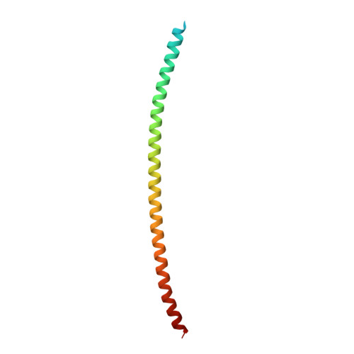
    HIV-1 and other enveloped viruses can be restricted by a host cellular protein called BST2/tetherin that prevents release of budded viruses from the cell surface. Mature BST2 contains a small cytosolic region, a predicted transmembrane helix, and an extracellular domain with a C-terminal GPI anchor. To advance understanding of BST2 function, we have determined a 2.6 Å crystal structure of the extracellular domain of the bacterially expressed recombinant human protein, residues 47-152, under reducing conditions. The structure forms a single long helix that associates as a parallel dimeric coiled coil over its C-terminal two-thirds, while the N-terminal third forms an antiparallel four-helix bundle with another dimer, creating a global tetramer. We also report the 3.45 Å resolution structure of BST2(51-151) prepared by expression as a secreted protein in HEK293T cells. This oxidized construct forms a dimer in the crystal that is superimposable with the reduced protein over the C-terminal two-thirds of the molecule, and its N terminus suggests pronounced flexibility. Hydrodynamic data demonstrated that BST2 formed a stable tetramer under reducing conditions and a dimer when oxidized to form disulfide bonds. A mutation that selectively disrupted the tetramer (L70D) increased protein expression modestly but only reduced antiviral activity by approximately threefold. Our data raise the possibility that BST2 may function as a tetramer at some stage, such as during trafficking, and strongly support a model in which the primary functional state of BST2 is a parallel disulfide-bound coiled coil that displays flexibility toward its N terminus.


  • Organizational Affiliation

    Department of Biochemistry, University of Utah School of Medicine, Salt Lake City, UT 84112-5650, USA.


Macromolecules
Find similar proteins by:  (by identity cutoff)  |  3D Structure
Entity ID: 1
MoleculeChains Sequence LengthOrganismDetailsImage
BONE MARROW STROMAL ANTIGEN 2A,
B [auth C]
103Homo sapiensMutation(s): 0 
UniProt & NIH Common Fund Data Resources
Find proteins for Q10589 (Homo sapiens)
Explore Q10589 
Go to UniProtKB:  Q10589
PHAROS:  Q10589
GTEx:  ENSG00000130303 
Entity Groups  
Sequence Clusters30% Identity50% Identity70% Identity90% Identity95% Identity100% Identity
UniProt GroupQ10589
Sequence Annotations
Expand
  • Reference Sequence
Small Molecules
Ligands 1 Unique
IDChains Name / Formula / InChI Key2D Diagram3D Interactions
NAG
Query on NAG

Download Ideal Coordinates CCD File 
C [auth A],
D [auth C]
2-acetamido-2-deoxy-beta-D-glucopyranose
C8 H15 N O6
OVRNDRQMDRJTHS-FMDGEEDCSA-N
Experimental Data & Validation

Experimental Data

  • Method: X-RAY DIFFRACTION
  • Resolution: 3.45 Å
  • R-Value Free: 0.298 
  • R-Value Work: 0.269 
  • R-Value Observed: 0.270 
  • Space Group: P 21 21 21
Unit Cell:
Length ( Å )Angle ( ˚ )
a = 28.86α = 90
b = 91.86β = 90
c = 146.96γ = 90
Software Package:
Software NamePurpose
REFMACrefinement
MOSFLMdata reduction
SCALAdata scaling
PHASERphasing

Structure Validation

View Full Validation Report


Ligand Structure Quality Assessment 


Entry History 

Deposition Data

Revision History  (Full details and data files)

  • Version 1.0: 2010-10-13
    Type: Initial release
  • Version 1.1: 2011-05-08
    Changes: Version format compliance
  • Version 1.2: 2011-07-13
    Changes: Version format compliance
  • Version 1.3: 2020-07-29
    Type: Remediation
    Reason: Carbohydrate remediation
    Changes: Data collection, Derived calculations, Other, Structure summary
  • Version 1.4: 2023-12-20
    Changes: Data collection, Database references, Refinement description, Structure summary